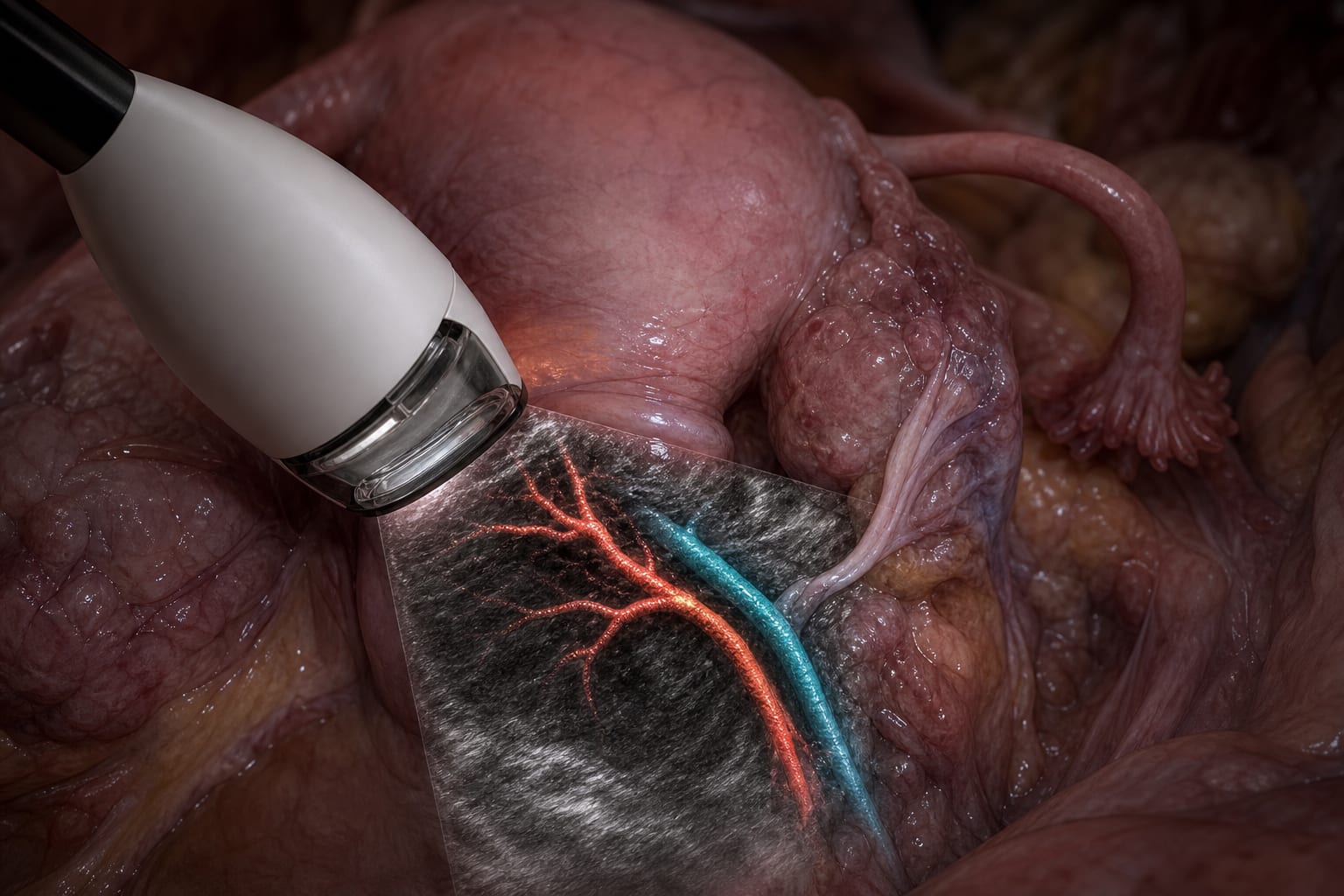
Clinical Scorecard: Ferroptosis Induced by Iron Overload in CD8+ T Cells Causes Functional Impairments That Accelerate Endometriosis Progression
At a Glance
Category
Detail
Condition
Endometriosis (EM)
Key Mechanisms
Iron overload induces ferroptosis in CD8+ T cells, impairing their function and accelerating EM progression
Target Population
Women of reproductive age with stage III–IV endometriosis
Care Setting
Specialist gynecology and immunology research settings
Key Highlights
EM lesions exhibit localized iron overload due to periodic bleeding of ectopic endometrial tissue.
Excess iron triggers reactive oxygen species generation leading to lipid peroxidation and ferroptosis.
Ferroptosis in CD8+ T cells diminishes their activation and cytotoxicity, impairing lesion clearance.
Guideline-Based Recommendations
Diagnosis
Confirm EM diagnosis via ultrasound and histopathology.
Assess iron overload and immune cell infiltration in eutopic and ectopic endometrial tissues.
Management
Consider interventions targeting iron overload and ferroptosis pathways to restore CD8+ T cell function.
Adoptive transfer of functional CD8+ T cells may reduce lesion burden.
Monitoring & Follow-up
Monitor CD8+ T cell activation status and ferroptosis markers in EM lesions.
Evaluate iron levels and oxidative stress markers in affected tissues.
Risks
Iron overload may exacerbate inflammatory microenvironment and promote EM progression.
Ferroptosis-induced CD8+ T cell dysfunction can impair immune surveillance and lesion clearance.
Patient & Prescribing Data
Women aged 20–49 years with regular menstrual cycles and stage III–IV EM
No direct prescribing data; potential therapeutic strategies include modulation of iron levels and ferroptosis inhibition to restore CD8+ T cell function.
Clinical Best Practices
Use minimally invasive tissue sampling for immunohistochemistry and flow cytometry analyses.
Employ in vitro and in vivo models to evaluate ferroptosis-targeted interventions.
Ensure ethical compliance and humane treatment in animal and human studies.
These 10 states make it more practical for physicians to participate in hospital ownership by aligning statutory structure, corporate practice of medicine rules, and population trends.
A VHA study across 11 vendors finds AI-generated primary care notes score lower than clinician-written notes, with the largest deficits in thoroughness, organization, and usefulness