Unsupervised anomaly detection for longitudinal comparison in whole-body PET/CT images
-
By
-
Takahiro Nakao
-
Shouhei Hanaoka
-
Yukihiro Nomura
-
Takeharu Yoshikawa
-
Osamu Abe
-
May 25, 2026
-
Clinical Scorecard: Automated Anomaly Identification for Longitudinal Analysis in Whole-Body PET/CT Imaging
At a Glance
| Category | Detail |
|---|
| Condition | Longitudinal comparison of whole-body PET/CT imaging |
| Key Mechanisms | Unsupervised anomaly detection to identify newly appearing lesions |
| Target Population | Adults undergoing whole-body medical screening |
| Care Setting | Hospital-based imaging facilities |
Key Highlights
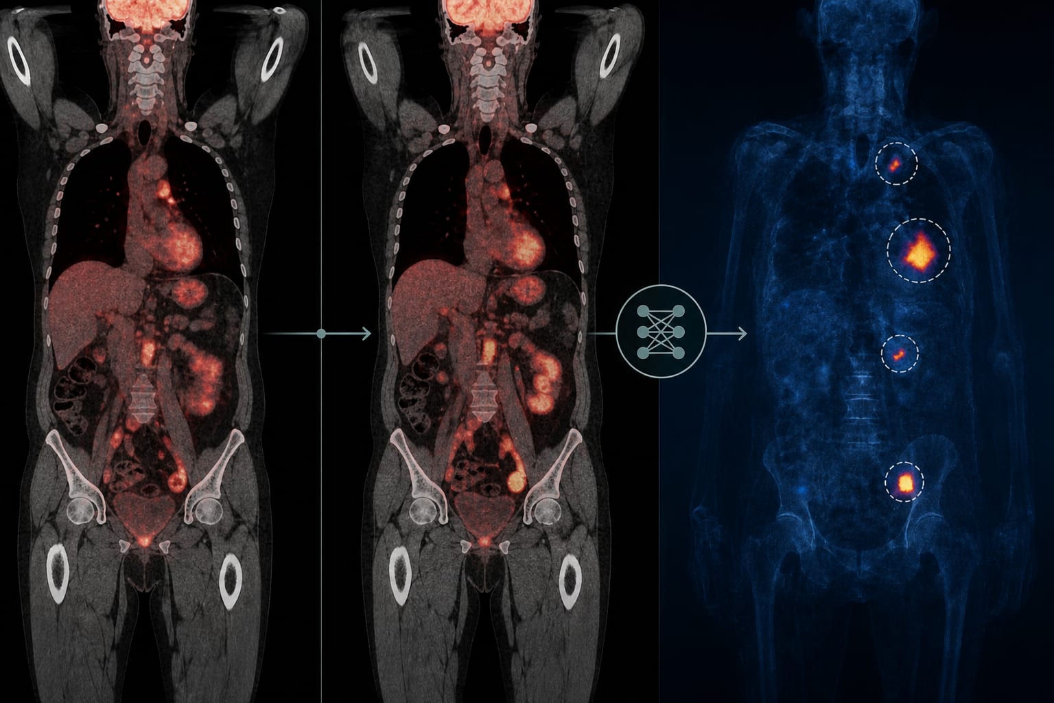
- Longitudinal comparison reduces false positives compared to subtraction methods
- Unsupervised anomaly detection does not require lesion annotations
- Method captures arbitrary types of abnormalities across the whole body
- Study involved 4,176 subjects with multiple PET/CT examinations
- Final diagnosis determined by consensus of two radiologists
Guideline-Based Recommendations
Diagnosis
- Use double-reading approach for interpreting PET/CT images
- Classify images as abnormal or normal based on FDG uptake
Management
- Further diagnostic evaluation or treatment at a referral center for abnormal findings
Monitoring & Follow-up
- Perform PET/CT examinations at approximately 1-year intervals
Risks
- False positives due to image misregistration and physiological tracer uptake variability
Patient & Prescribing Data
Adults with normal and abnormal PET/CT findings
Focus on newly diagnosed abnormalities during follow-up
Clinical Best Practices
- Implement unsupervised anomaly detection for improved lesion identification
- Ensure thorough training and validation datasets for model development
Related Resources & Content