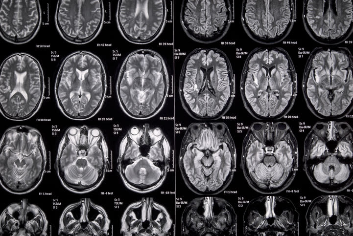
Meningococcal Abscess Detected by MRI
MRI identifies rare intracranial complication following normal computed tomography
-
By
-
Julie Greenbaum
-
March 27, 2026
-
Clinical Scorecard: Meningococcal Abscess Detected by MRI
At a Glance
| Category | Detail |
|---|
| Condition | Meningococcal meningitis with cerebellar abscess |
| Key Mechanisms | Detection of cerebellar abscess via MRI despite normal CT scan |
| Target Population | Pediatric patients, specifically older children |
| Care Setting | Intensive care unit and outpatient follow-up |
Key Highlights
- Cerebellar abscess detected only by MRI in a child with meningococcal meningitis
- Initial CT scan was unremarkable; MRI revealed focal changes consistent with abscess
- Patient improved with antimicrobial therapy and was extubated by day 3
- Multidisciplinary team recommended a 21-day course of intravenous ceftriaxone
- Follow-up MRI showed resolution of the cerebellar abscess by day 26
Guideline-Based Recommendations
Diagnosis
- Use MRI for early detection of parenchymal infection in atypical meningitis cases
Management
- Administer targeted antimicrobial therapy based on confirmed diagnosis
- Nonoperative management recommended for small lesions without mass effect
Monitoring & Follow-up
- Serial MRI to monitor lesion progression and guide treatment
Risks
- Brain abscess occurs in fewer than 1% of invasive meningococcal infections
Patient & Prescribing Data
Previously healthy children with meningococcal meningitis
Early recognition and targeted therapy can lead to favorable outcomes
Clinical Best Practices
- Maintain heightened clinical suspicion for atypical neurological findings in meningitis
- Utilize MRI for monitoring and diagnosis when CT results are inconclusive
References