Clinical Scorecard: Obstacles in the Segmentation of Intraoperative Ultrasound Images for Brain Tumor Surgery
At a Glance
Category
Detail
Condition
Brain tumors requiring maximal-safe resection
Key Mechanisms
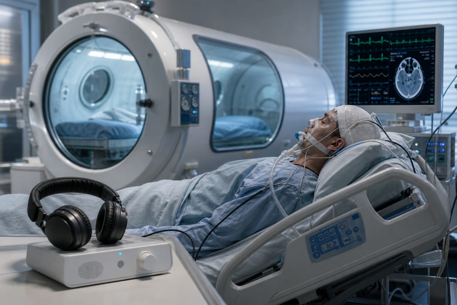
Intraoperative ultrasound (iUS) imaging for real-time tumor detection and delineation
Target Population
Patients undergoing brain tumor surgery with intraoperative ultrasound guidance
Care Setting
Neurosurgical operating room with intraoperative imaging
Key Highlights
iUS offers real-time tumor visualization integrated into surgical workflow and is more affordable than intraoperative MRI.
Challenges in iUS include limited field of view, artefacts, steep learning curve, and variability in tumor appearance and intraoperative changes.
Current evidence shows iUS has moderate sensitivity (72.2%) and high specificity (93.5%) for glioma resection assessment but requires accuracy improvements.
Guideline-Based Recommendations
Diagnosis
Use iUS co-registered with preoperative MRI/CT for intraoperative tumor boundary delineation.
Cross-reference iUS images with preoperative MRI to ensure accurate tumor boundary identification.
Management
Employ iUS to guide maximal-safe tumor resection to improve symptoms, quality of life, and survival.
Incorporate standardized training and new supporting techniques to reduce segmentation errors and improve iUS utility.
Monitoring & Follow-up
Assess tumor resection completeness intraoperatively using iUS with reference to postoperative MRI.
Monitor interobserver variability in tumor boundary segmentation to identify areas needing training or tool improvement.
Risks
Potential for incomplete tumor resection or inadvertent damage due to inaccurate tumor boundary detection on iUS.
Steep learning curve and variability in image interpretation may impair surgical outcomes.
Patient & Prescribing Data
Patients with brain tumors undergoing iUS-guided resection
iUS-guided resection achieves approximately 77% gross total resection rate, comparable to other navigation methods, but accuracy improvements are needed for standard care adoption.
Clinical Best Practices
Utilize experienced operators and standardized protocols for iUS image acquisition and interpretation.
Combine iUS with preoperative MRI for improved tumor boundary delineation.
Implement training programs to overcome the steep learning curve and improve segmentation consistency.
Explore simplified annotation methods such as bounding boxes to complement detailed segmentation and reduce variability.