Neurofilament light and glial fibrillary acidic protein do not reflect neuronal or glial damage during different intracranial radiotherapy regimes: a pilot study - Scorecard - MDSpire
Advertisement
Neurofilament light and glial fibrillary acidic protein do not reflect neuronal or glial damage during different intracranial radiotherapy regimes: a pilot study
Clinical Scorecard: Neurofilament Light Chain and Glial Fibrillary Acidic Protein Levels Do Not Indicate Neuronal or Glial Injury Across Various Intracranial Radiotherapy Protocols: A Preliminary Investigation
At a Glance
Category
Detail
Condition
Key Mechanisms
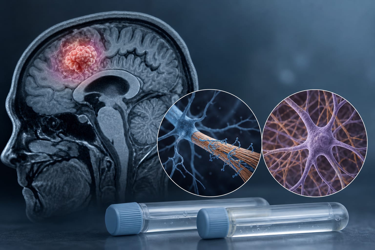
Neurofilament light chain (NfL) and glial fibrillary acidic protein (GFAP) as non-specific markers of central nervous system damage.
Target Population
Care Setting
Key Highlights
Elevated NfL and GFAP levels observed before radiotherapy initiation.
No significant increase in NfL and GFAP levels during radiotherapy.
Decreasing NfL and GFAP values correlated with treatment response during follow-up.
Pronounced increases in NfL levels were associated with new cerebral lesions.
Guideline-Based Recommendations
Diagnosis
Monitor NfL and GFAP levels.
Management
Monitoring & Follow-up
Utilize NfL and GFAP levels for follow-up assessments post-treatment.
Risks
Increased NfL levels may indicate new cerebral lesions.
Patient & Prescribing Data
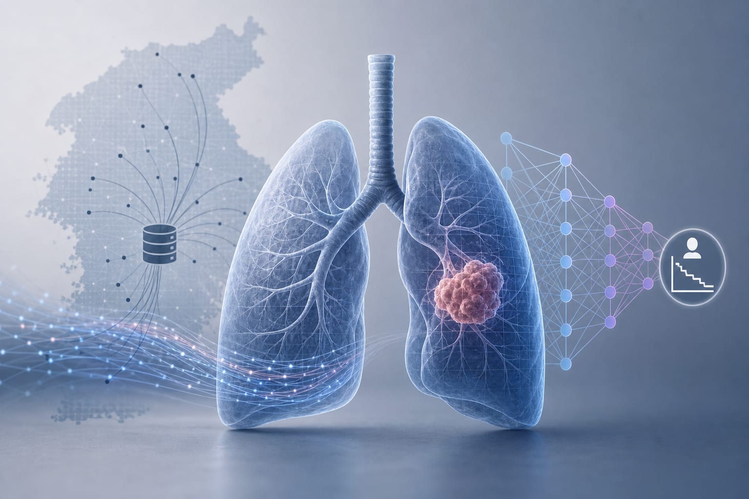
Stereotactic radiotherapy (SRT) shows high local control rates.
by Yvonne Dzierma, Holger Sebb, Michael Utzig, Nurlan Abdullayev, Christian Berdel, Christian Ruebe, Jochen Fleckenstein, Markus Hecht, Guido Hildebrandt, Mathias Jucker, Kristina Heyne