Paucibacillary Tuberculous Meningitis: Navigating the Diagnostic Difficulties of Clinical-Radiological Discrepancies
-
By
-
Tianjiao Tang
-
Junguo Cao
-
Mantao Chen
-
Yao Yao
-
Luqing Tong
-
September 3, 2026
Clinical Scorecard: Paucibacillary Tuberculous Meningitis: Navigating the Diagnostic Difficulties of Clinical-Radiological Discrepancies
At a Glance
Category | Detail |
|---|---|
Condition | Paucibacillary tuberculous meningitis |
Key Mechanisms | Diagnosis may be complicated by atypical clinical features, near-normal CSF findings, and negative microbiological and molecular tests. |
Target Population | This case involved a 63-year-old woman with newly diagnosed diabetes who presented with acute neurologic symptoms but lacked typical infectious features of TBM. |
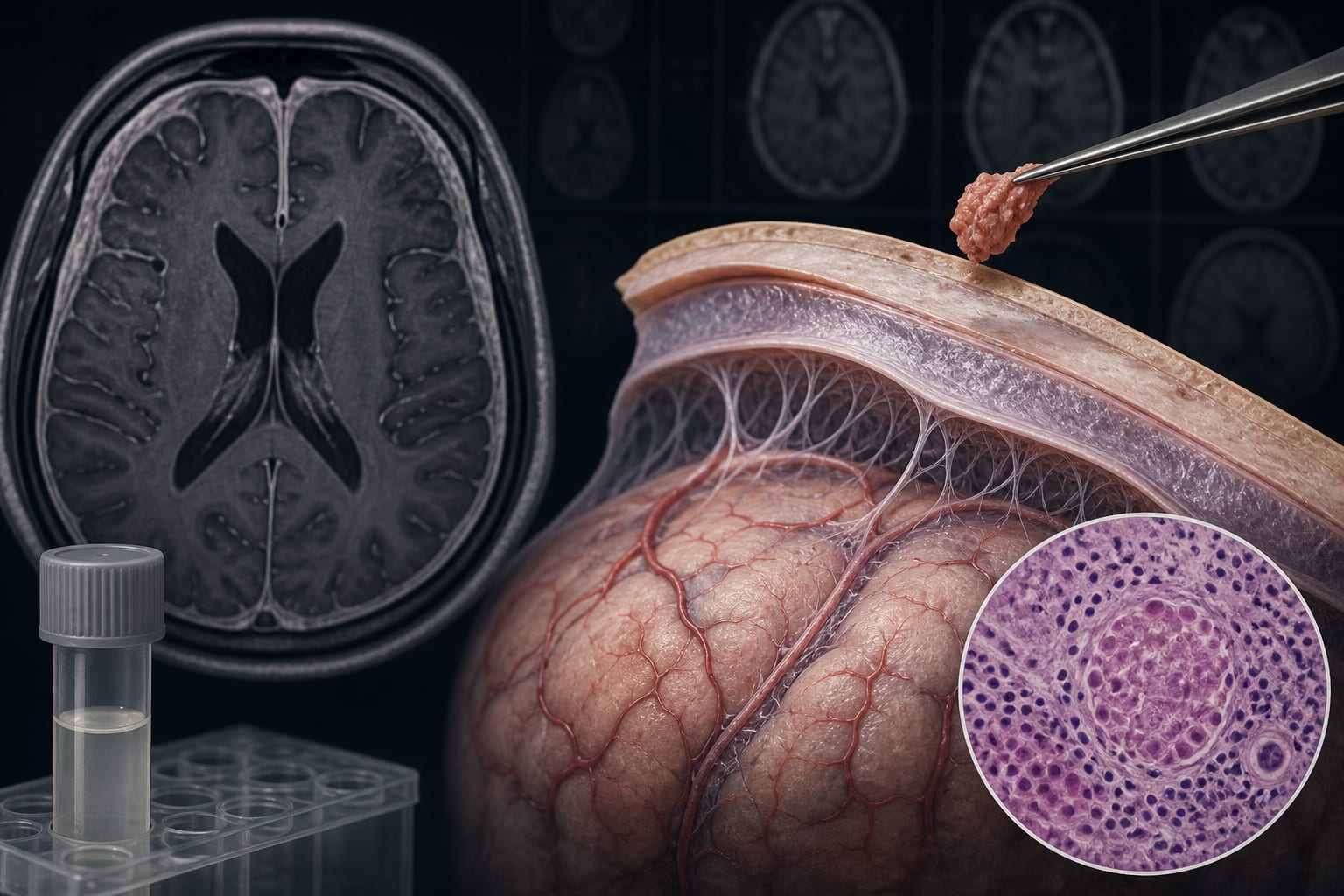
Care Setting | Neurologic and infectious disease diagnostic evaluation, including meningeal biopsy after clinical improvement was accompanied by radiological progression. |
Key Highlights
TBM mortality exceeds 20% even with appropriate treatment.
The Marais criteria provide a standardized framework for clinical research, with a score of ≥6 required for possible TBM.
Normal or near-normal CSF findings can occur in TBM and do not exclude paucibacillary disease.
Meningeal biopsy showed granulomatous inflammation, and a single acid-fast bacillus was identified; together with a positive IGRA, these findings strongly supported a tuberculous etiology.
Follow-up MRI after 9 months showed almost complete resolution of meningeal enhancement and cerebral edema, and the patient achieved full neurological recovery.
Guideline-Based Recommendations
Diagnosis
The Marais criteria provide a standardized framework for TBM classification in clinical research; in this patient, the initial score was 2, below the threshold for possible TBM.
Negative CSF GeneXpert MTB/RIF, mycobacterial culture, and metagenomic next-generation sequencing did not exclude paucibacillary TBM in this case.
The authors state that radiological progression despite clinical quiescence should prompt strong consideration of meningeal biopsy, particularly when molecular tests on CSF remain negative.
Management
In this case, anti-tuberculosis treatment lasted approximately 12 months and included isoniazid, rifampicin, pyrazinamide, moxifloxacin, and linezolid.
Dexamethasone was administered for more than 3 months and gradually tapered.
Monitoring & Follow-up
During the documented 9-month treatment period, the patient attended monthly follow-up visits, maintained excellent adherence, and experienced no significant adverse events.
Follow-up MRI after 9 months demonstrated almost complete resolution of meningeal enhancement and cerebral edema, and the patient achieved full neurological recovery.
Risks
Complete symptomatic improvement may occur despite radiological progression and does not necessarily indicate disease control.
The authors caution that a transient clinical response to corticosteroids without specific anti-tuberculosis therapy may create a false sense of security and delay definitive diagnosis and treatment.
Patient & Prescribing Data
The patient was a 63-year-old woman with newly diagnosed diabetes who presented with sudden-onset headache, dysarthria, right-sided weakness, and two focal seizures. She was afebrile, lacked meningeal irritation signs, and denied preceding fever, cough, or respiratory symptoms.
Anti-tuberculosis therapy lasted approximately 12 months and included isoniazid, rifampicin, pyrazinamide, moxifloxacin, and linezolid. Dexamethasone was administered for more than 3 months.
Clinical Best Practices
Maintain clinical suspicion for TBM when patients have atypical neurologic presentations despite negative microbiological testing.
Recognize that normal or near-normal CSF findings do not exclude paucibacillary TBM.
Consider meningeal biopsy when radiological progression occurs despite clinical improvement and molecular tests on CSF remain negative.
Interpret a positive IGRA together with histopathological and clinical findings; in this case, the acid-fast bacillus and positive IGRA strongly supported tuberculous etiology.
Related Resources & Content
Heemskerk AD, Bang ND, Mai NT, Chau TT, Phu NH, Loc PP, et al. “Intensified antituberculosis therapy in adults with tuberculous meningitis.” New England Journal of Medicine. 2016;374(2):124–134.
Wilkinson RJ, Rohlwink U, Misra UK, van Crevel R, Thwaites GE. “Tuberculous meningitis.” Nature Reviews Neurology. 2017;13(10):581–598.
Leiva-Ordoñez JE, Quintero B. “Diagnosis of tuberculous meningitis: integrating clinical assessment and molecular diagnostics.” Diagnostics. 2026;16(4):552.
Marais S, Thwaites G, Schoeman JF, Trk ME, Marais BJ. “Tuberculous meningitis: a uniform case definition for use in clinical research.” The Lancet Infectious Diseases. 2010;10(11):803–812.
Bahr NC, Nuwagira E, Evans EE, Cresswell FV, Bystrom PV, Byamukama A, et al. “Diagnostic accuracy of Xpert MTB/RIF Ultra for tuberculous meningitis in HIV-infected adults: a prospective cohort study.” The Lancet Infectious Diseases. 2018;18(1):68–75.
Thwaites GE, Nguyen DB, Nguyen HD, Hoang TQ, Do TT, Nguyen TC, et al. “Dexamethasone for the treatment of tuberculous meningitis in adolescents and adults.” New England Journal of Medicine. 2004;351(17):1741–1751.
Based on findings from:
Paucibacillary tuberculous meningitis with clinical-radiological dissociation: A diagnostic challenge
Tianjiao Tang, Junguo Cao, Mantao Chen, Yao Yao, Luqing Tong. International Journal Of Infectious Diseases, 2026.
https://www.sciencedirect.com/science/article/pii/S1201971226006983
This content is an AI-generated, fully rewritten summary based on a published scholarly article. It does not reproduce the original text and is not a substitute for the original publication. Readers are encouraged to consult the source for full context, data, and methodology.