Imaging-based techniques for ablation zone definition and volumetry after laser interstitial thermal therapy (LITT) for intracranial lesions: a systematic review - Scorecard - MDSpire
Advertisement
Imaging-based techniques for ablation zone definition and volumetry after laser interstitial thermal therapy (LITT) for intracranial lesions: a systematic review
Clinical Scorecard: Techniques Utilizing Imaging for Defining Ablation Zones and Assessing Volume Post-Laser Interstitial Thermal Therapy for Intracranial Lesions: A Comprehensive Review
At a Glance
Category
Detail
Condition
Intracranial lesions including high-grade gliomas, epilepsy, and radiation necrosis
Key Mechanisms
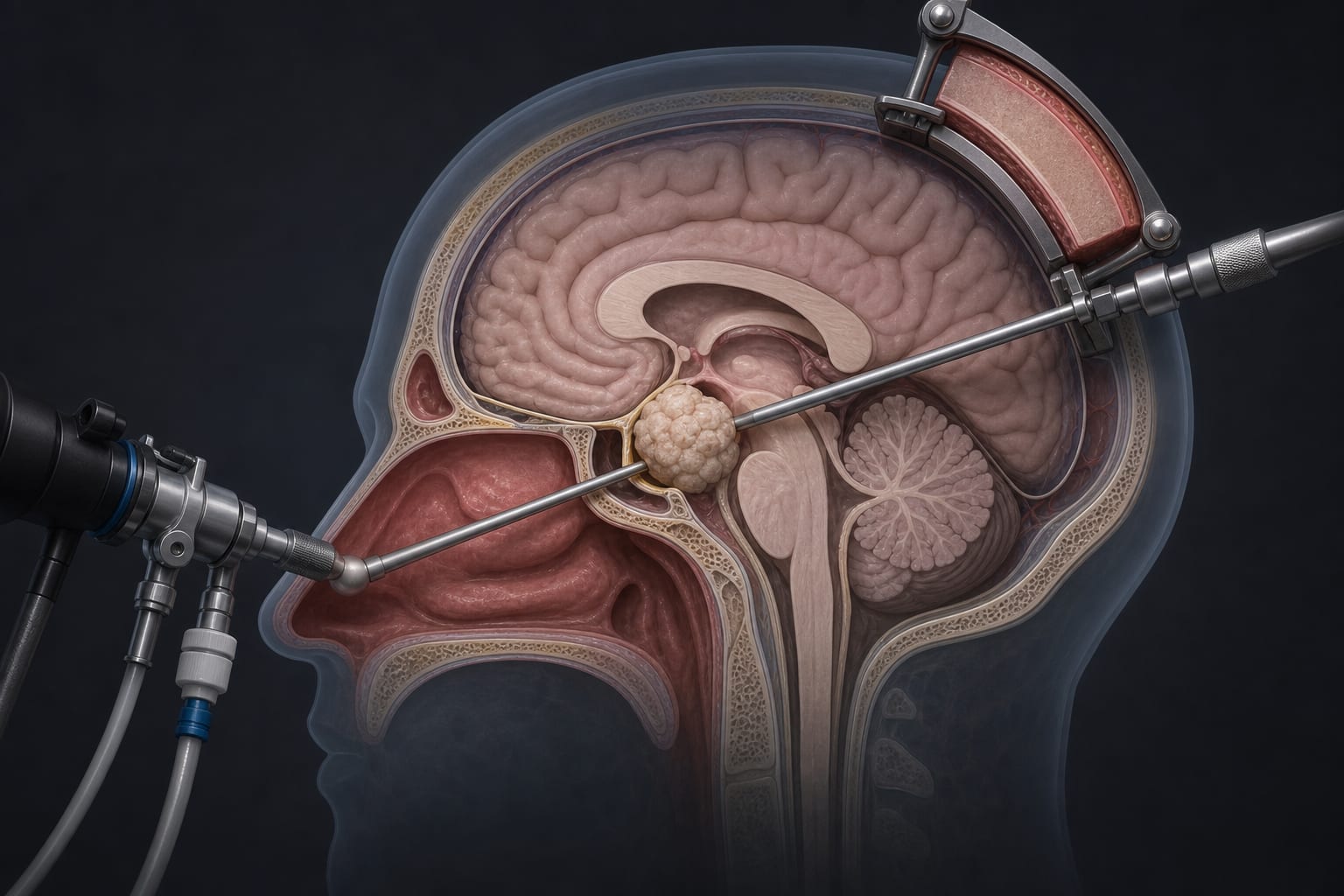
Laser interstitial thermal therapy (LITT) uses MRI-guided laser ablation to target and destroy pathological brain tissue with real-time MR-thermometry monitoring
Target Population
Patients with deep-seated or eloquent-area brain lesions such as basal ganglia tumors or hypothalamic hamartomas
Care Setting
Neurosurgical and neuro-oncological centers equipped with MRI-guided laser ablation technology
Key Highlights
LITT enables minimally invasive cytoreduction of intracranial lesions in challenging locations previously inaccessible to surgery
Extent of ablation (EOA) correlates with prognosis but lacks standardized imaging-based volumetric assessment methods
Post-LITT ablation zones are visualized on contrast-enhanced T1-weighted MRI as a necrotic core with a peripheral enhancing rim, which can be difficult to distinguish from residual tumor
Guideline-Based Recommendations
Diagnosis
Use MRI guidance and real-time MR-thermometry during LITT to accurately target lesions
Perform immediate post-procedural contrast-enhanced T1-weighted MRI to visualize ablation zones
Management
Tailor ablation volume intraoperatively to maximize lesion cytoreduction while preserving surrounding healthy brain tissue
Consider LITT for patients with deep-seated or eloquent-area intracranial lesions unsuitable for open surgery
Monitoring & Follow-up
Assess ablation volume and extent of ablation using imaging modalities post-LITT
Recognize challenges in differentiating ablation zone rim enhancement from residual tumor on MRI
Use thermometry data and surgeon estimates as adjuncts for ablation volume assessment
Risks
Potential damage to surrounding healthy brain tissue if ablation is not precisely controlled
Difficulty in accurately delineating ablation zones may affect assessment of treatment efficacy
Patient & Prescribing Data
Patients undergoing LITT for intracranial lesions including tumors and epilepsy-related lesions
LITT offers a minimally invasive option with real-time monitoring to optimize ablation extent; however, standardized imaging protocols for post-treatment volume assessment are lacking
Clinical Best Practices
Employ MRI guidance with real-time thermometry during LITT for precise ablation targeting
Perform immediate post-LITT contrast-enhanced MRI to evaluate ablation zones
Use a combination of imaging, thermometry data, and clinical judgment to estimate extent of ablation
Recognize limitations in current imaging methods and advocate for development of standardized volumetric assessment protocols