Clinical Scorecard: Prediction of Ischemic Stroke in Cervical Artery Dissection Using Clinical Data and High-Resolution Magnetic Resonance Imaging Techniques
At a Glance
Category
Detail
Condition
Cervical Artery Dissection (CeAD)
Key Mechanisms
Intraluminal thrombus, white blood cell count, severe stenosis or occlusion, and alcohol consumption are associated with ischemic events.
Target Population
Patients with cervical artery dissection, particularly young and middle-aged adults.
Care Setting
Clinical evaluation and management of patients with suspected cerebrovascular events.
Key Highlights
CeAD accounts for 8-25% of ischemic strokes in individuals under 50 years.
More than 50% of CeAD patients may develop ischemic stroke or transient ischemic attack.
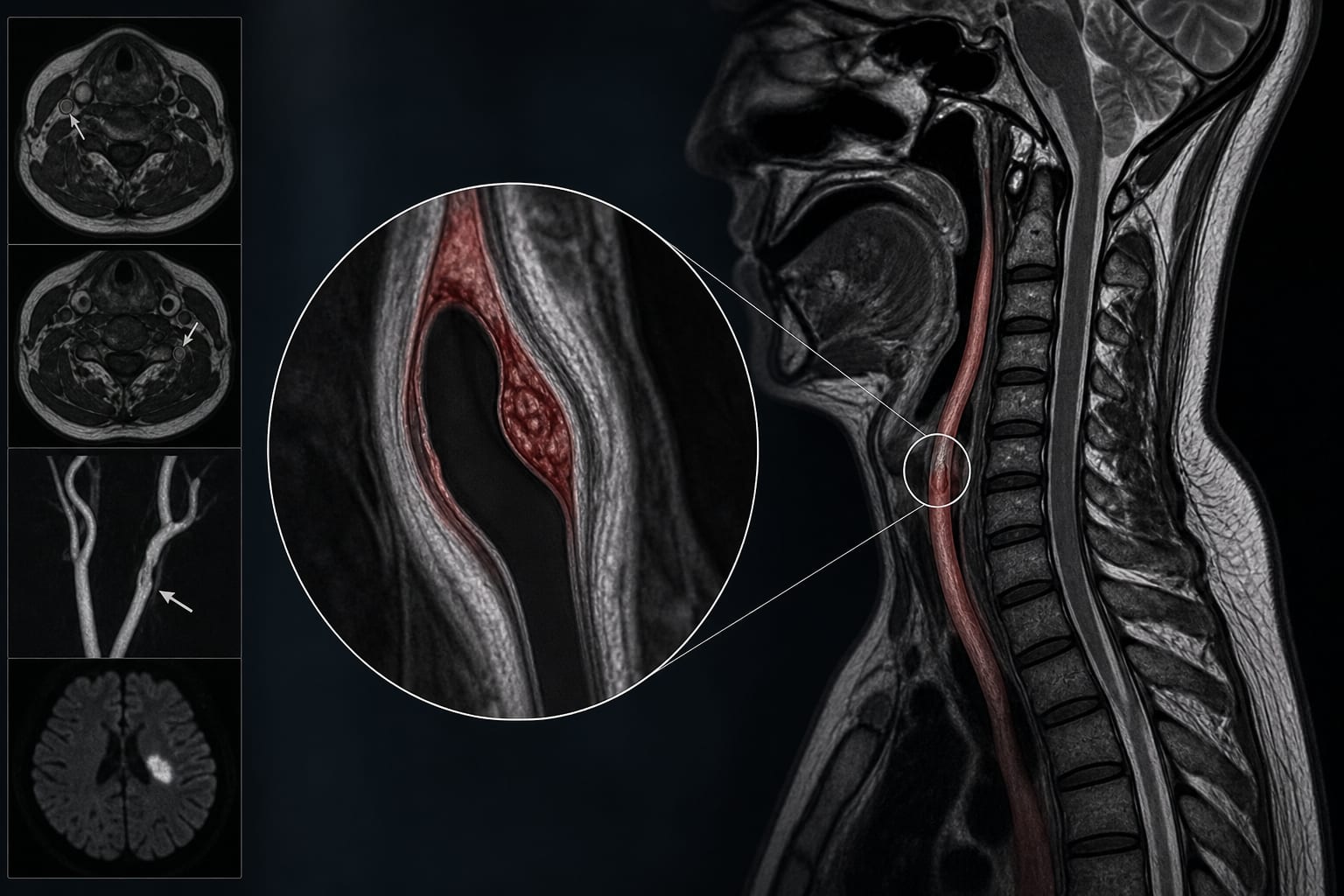
High-resolution magnetic resonance imaging (HRMRI) is crucial for diagnosis and risk stratification.
Guideline-Based Recommendations
Diagnosis
Diagnosis of CeAD should be based on HRMRI findings demonstrating intramural hematoma.
Management
Early identification of high-risk patients is essential for optimizing clinical management.
Monitoring & Follow-up
Patients should be monitored for ischemic events within the first 2 weeks after diagnosis.
Risks
Risk factors for ischemic stroke include WBC count, intraluminal thrombus, male sex, and alcohol consumption.
Patient & Prescribing Data
129 patients with cervical artery dissection.
A nomogram integrating clinical and imaging features shows good performance in predicting ischemic stroke risk.
Clinical Best Practices
Utilize HRMRI for detailed visualization of arterial wall and lumen in CeAD.
Incorporate both clinical data and imaging features for individualized risk prediction.