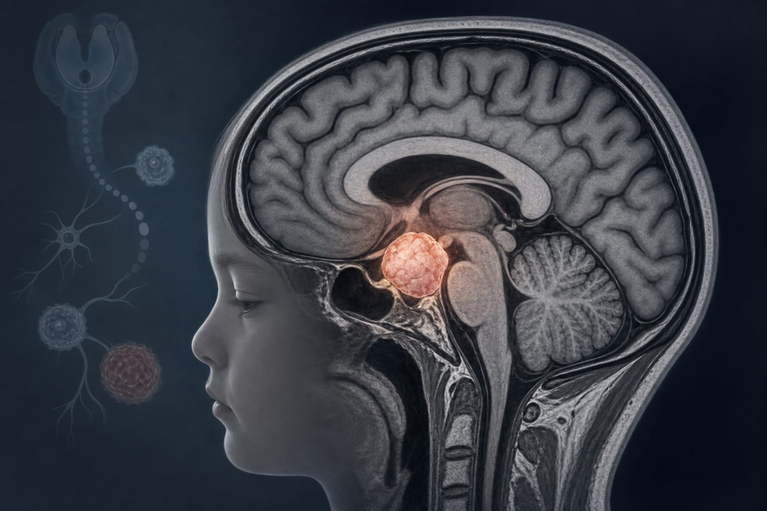
Clinical Scorecard: Clinical and Imaging Features of Intracerebral Hemorrhage Associated with Brain Tumors and Metastatic Lesions
At a Glance
Category
Detail
Condition
Neoplastic intracerebral hemorrhage (ICH) arising from brain tumors and metastases
Key Mechanisms
Rhexis bleeding of fragile neo-angiogenetic tumor vessels within neoplastic brain tumors or metastases
Target Population
Patients with primary brain tumors or metastatic brain lesions presenting with acute intracerebral hemorrhage
Care Setting
Neuro-oncology and stroke care units in tertiary care hospitals
Key Highlights
Neoplastic ICH is clinically challenging to distinguish from hypertensive or other non-neoplastic ICH, leading to potential delays in diagnosis and treatment.
Distinct clinical and radiographic features may differentiate ICH associated with primary brain tumors from metastatic lesions.
Quantitative imaging and detailed clinical characterization improve diagnostic accuracy and patient management.
Guideline-Based Recommendations
Diagnosis
Use the H-ATOMIC Classification system to categorize etiology of occult acute ICH.
Exclude other causes such as hypertension, cerebral amyloid angiopathy, coagulopathies, vascular malformations, trauma, and hemorrhagic transformation post-ischemic stroke.
Employ advanced imaging including CT and follow-up MRI to detect underlying neoplastic lesions masked by hemorrhage.
Management
Consider prior therapies such as craniotomy, chemo-, immunotherapy, radiotherapy, and steroid treatment in management planning.
Monitor and manage vascular risk factors and pre-existing anticoagulation or antiplatelet therapies.
Tailor treatment based on tumor type (primary vs metastatic) and hemorrhage characteristics.
Monitoring & Follow-up
Assess Glasgow Coma Scale (GCS) at admission and modified Rankin Scale (mRS) at discharge or last evaluation to monitor neurological status and functional outcome.
Perform volumetric quantification of ICH and perihematomal edema on imaging.
Evaluate for secondary intraventricular hemorrhage and hemorrhage location to guide prognosis and treatment.
Risks
Delayed diagnosis due to difficulty distinguishing neoplastic ICH from other hemorrhagic etiologies.
High mortality associated with neoplastic ICH.
Potential masking of underlying tumor by hemorrhage on initial imaging.
Patient & Prescribing Data
Patients with intracerebral hemorrhage secondary to primary brain tumors or metastatic brain lesions
Prior use of anticoagulants, antiplatelet agents, and therapies such as steroids, chemo-, immuno-, and radiotherapy influence clinical presentation and management strategies.
Clinical Best Practices
Perform comprehensive clinical assessment including neurological deficits, seizures, cognitive impairment, and systemic symptoms.
Use detailed imaging protocols to localize hemorrhage and identify tumor type.
Dichotomize mRS scores to evaluate functional independence versus need for assistance.
Integrate clinical, imaging, and treatment history for accurate diagnosis and timely intervention.
by Semil Eminovic, Tobias Orth, Andrea Dell’Orco, Lukas Baumgärtner, Andrea Morotti, David Wasilewski, Melisa S. Guelen, Michael Scheel, Tobias Penzkofer, Jawed Nawabi