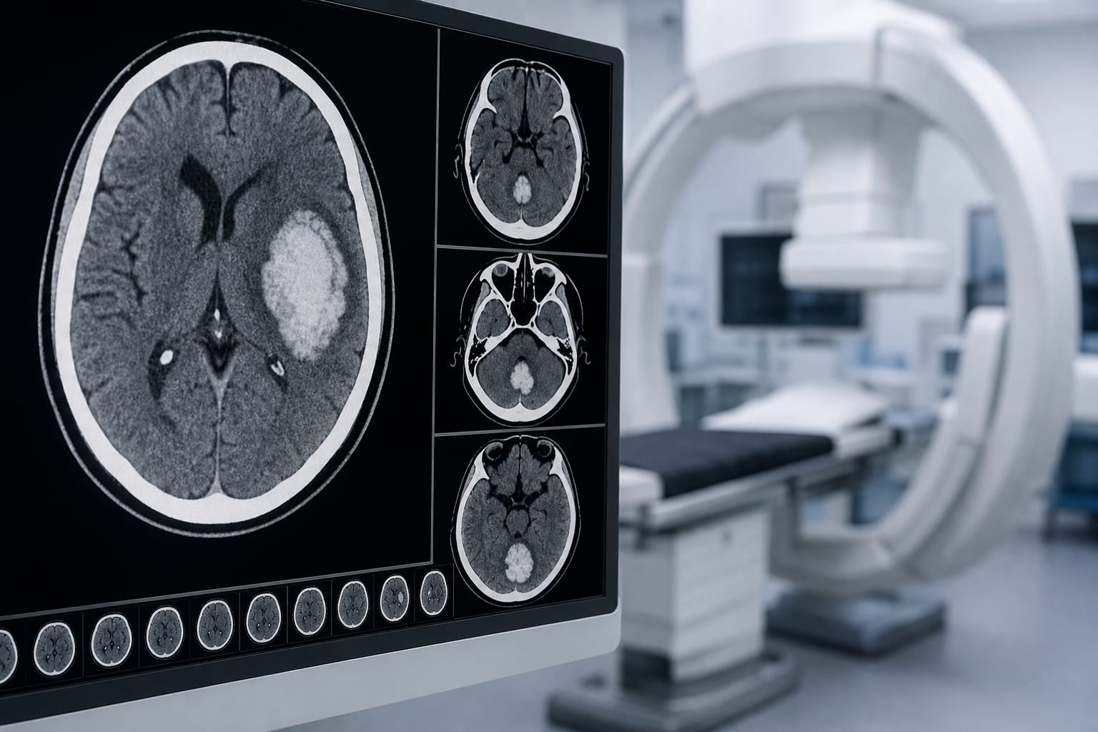
Size and location of intracranial hemorrhage in acute stroke presentations: correlation with baseline NIHSS and relevance to direct-to-angiography workflows - Scorecard - MDSpire
Conexiant’s news site is now MDSpire News. Learn more
Advertisement
Assessment of Intracranial Hemorrhage Size and Location in Acute Stroke Cases: Its Relationship with Initial NIHSS Scores and Implications for Direct-to-Angiography Protocols
Clinical Scorecard: Assessment of Intracranial Hemorrhage Size and Location in Acute Stroke Cases: Its Relationship with Initial NIHSS Scores and Implications for Direct-to-Angiography Protocols
At a Glance
Category
Detail
Condition
Intracranial Hemorrhage in Acute Stroke
Key Mechanisms
Relationship between hemorrhage volume, location, and stroke severity as measured by NIHSS.
Target Population
Adults aged ≥18 years with spontaneous intracranial hemorrhage.
Care Setting
High-volume comprehensive stroke center.
Key Highlights
Intraparenchymal hemorrhage comprised 89% of cases.
Median hemorrhage volume was 18 mL, with significant correlation to NIHSS scores.
Lobar hemorrhages had the greatest volumes, while infratentorial hemorrhages caused severe deficits despite smaller volumes.
Only 2.8% of patients with NIHSS ≥7 had isolated infratentorial hemorrhage.
FDCT image quality in the posterior fossa is problematic due to beam-hardening artifacts.
Guideline-Based Recommendations
Diagnosis
Use flat-panel detector CT (FDCT) to rule out hemorrhage before thrombectomy in suspected large vessel occlusion.
Management
Consider direct-to-angiography (DTA) protocols to reduce treatment delays in acute stroke cases.
Monitoring & Follow-up
Monitor NIHSS scores to assess stroke severity and potential hemorrhage impact.
Risks
Approximately 20% of patients with NIHSS ≥7 may have hemorrhagic stroke, necessitating careful imaging assessment.
Patient & Prescribing Data
Adults with confirmed spontaneous intracranial hemorrhage.
DTA workflows may improve treatment times but require reliable hemorrhage exclusion.
Clinical Best Practices
Ensure high-quality imaging of the posterior fossa during DTA workflows.
Utilize semi-automated segmentation for accurate hemorrhage volume assessment.
Baptist Health will bring together leading physicians, researchers and healthcare professionals from South Florida and across the country for the 14th Annual Baptist Health Miami Neuroscience Symposium, taking place Nov. 4-6, 2026, at Hilton Miami Dadeland and virtually via Zoom.