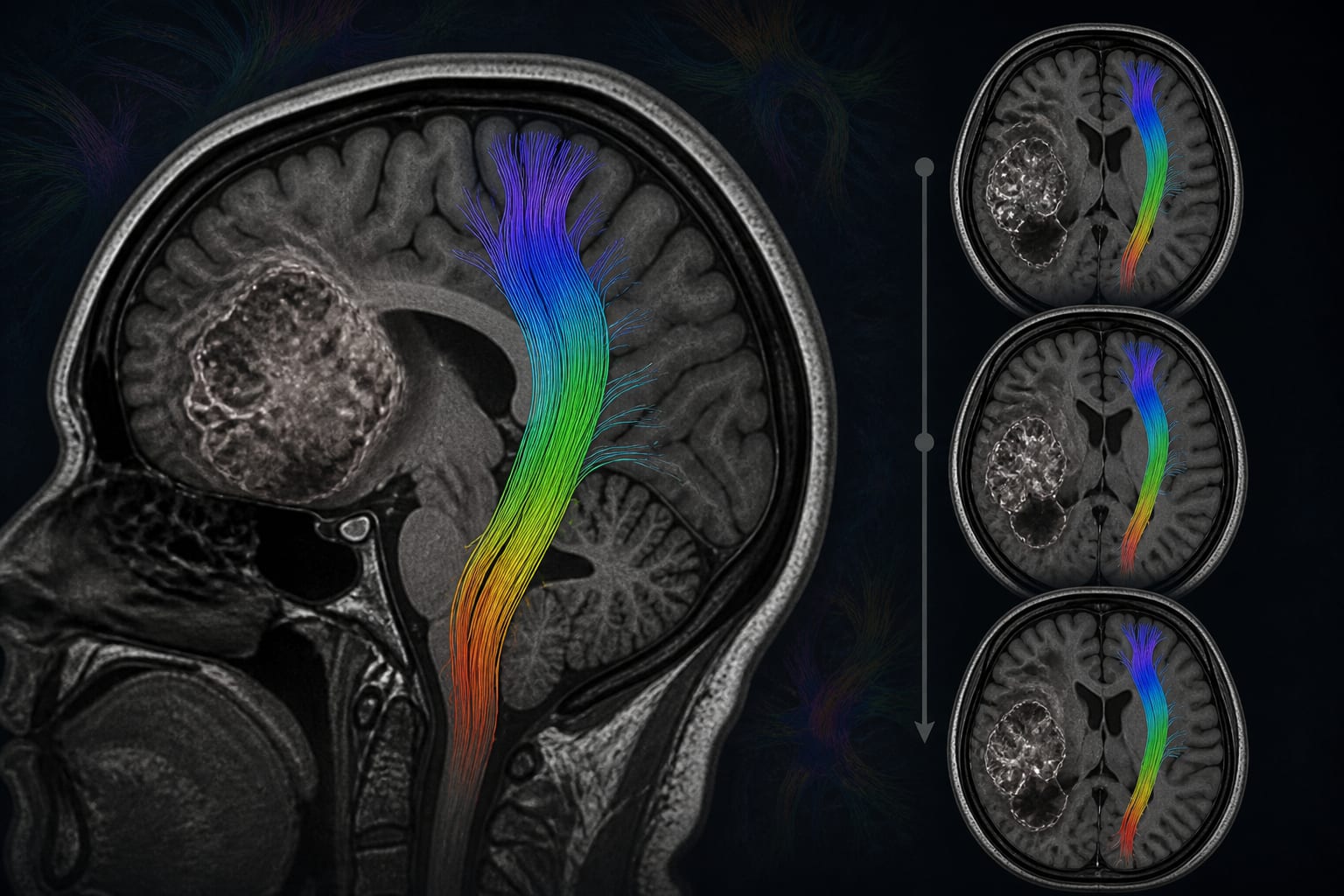
Longitudinal spatial changes of the contralesional corticospinal tract in high-grade glioma: a within-patient DTI tractography study - Scorecard - MDSpire
Clinical Scorecard: Within-Patient DTI Tractography Reveals Longitudinal Spatial Changes in the Contralesional Corticospinal Tract in High-Grade Glioma
At a Glance
Category
Detail
Condition
Clinical topic addressed by the source article: Within-Patient DTI Tractography Reveals Longitudinal Spatial Changes in the Contralesional Corticospinal Tract in High-Grade Glioma
Key Mechanisms
The introduction notes that gliomas may affect the CST through displacement, infiltration, disruption, or diffusion alterations. The study assessed tract-averaged fractional anisotropy, length, volume, trajectory, and voxel-level distribution.
Target Population
Patients or participants described in the source article.
Care Setting
Care setting described in the source article.
Key Highlights
The exploratory study compared contralesional CST measurements within patients at primary and recurrent high-grade glioma assessments.
The retrospective cohort comprised 29 adults treated from 2013 through 2021; all had MRC strength of 5/5 at both assessments.
Deterministic DTI-based tractography used the same anatomically guided reconstruction protocol at both assessments.
Spatial analysis normalized CST masks to MNI152 space and harmonized laterality for within-patient comparison.
The supplied article text describes the study methods but does not provide its results.
Guideline-Based Recommendations
Diagnosis
Study inclusion required clinically imaging-documented first recurrence.
Motor function was assessed clinically with the MRC scale; eligibility required strength of 5/5 at both assessments.
Management
Monitoring & Follow-up
Preoperative MRI and DTI were obtained 1–7 days before surgery at each assessment.
The same anatomically guided contralesional CST reconstruction protocol was used at primary and recurrent assessments.
Risks
The introduction notes that diffusion abnormalities have been described in contralateral normal-appearing white matter in malignant glioma.
The authors state that longitudinal contralesional CST variation, if present, may warrant caution when using the tract as a stable comparator in serial glioma research.
Patient & Prescribing Data
No prescribing or treatment-effect data are provided. The study cohort comprised 29 adults with frontal-lobe high-grade glioma and preserved MRC strength at both imaging assessments.
Treatment administered between assessments was recorded, but the supplied text does not report treatment outcomes or prescribing recommendations.
Clinical Best Practices
Study procedure: the reconstructed tractography-defined motor component of the CST was not the complete anatomical CST.
Study procedure: reconstructed streamlines underwent visual inspection, with anatomically implausible streamlines removed when necessary.
Study procedure: spatially transformed CST masks were checked for alignment with normalized T1-weighted images, and grid dimensions, affine information, and patient pairing were verified.
Study design: no direction of longitudinal change was prespecified because glioma-specific evidence was limited.
by Bingzhen Li, Raimunde Liang, Ghaith Altawalbeh, Lisa Hoenikl, Eugen Ursu, Axel Schroeder, Haosu Zhang, Philipp Karschnia, Chiara Negwer, Bernhard Meyer, Arthur Wagner
Harold Burstein, MD, PhD, and Ana C. Garrido-Castro, MD discuss new antibody drug conjugate treatment options in first line triple negative breast cancer.