Amazing Advancements in Anterior Segment Diagnostics
The latest on anterior segment OCT, corneal tomography, and confocal microscopy,
By
W. BARRY LEE, MD, FACS
June 1, 2025
Clinical Scorecard: Amazing Advancements in Anterior Segment Diagnostics
At a Glance
Category Detail
Condition Ocular surface and anterior segment diseases Key Mechanisms Utilization of advanced imaging technologies like AS-OCT and corneal tomography for diagnosis and management. Target Population Patients with anterior segment pathologies, including keratoconus and ocular surface tumors. Care Setting Ophthalmology clinics and surgical centers.
Key Highlights
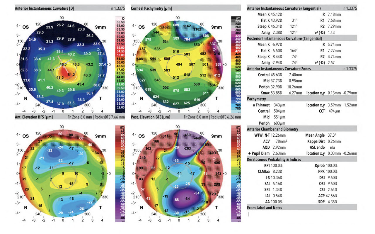
AS-OCT provides high-resolution imaging for anterior segment structures. Epithelial mapping aids in diagnosing keratoconus and tracking progression. Corneal tomography helps identify keratoconus risk in refractive surgery candidates. Confocal microscopy is crucial for diagnosing fungal and Acanthamoeba keratitis. Advanced metrics enhance the evaluation of corneal diseases. Guideline-Based Recommendations
Diagnosis
Use AS-OCT for assessing corneal pathology and ocular surface tumors. Employ corneal tomography for keratoconus screening. Management
Consider phototherapeutic keratectomy for superficial corneal pathology. Utilize deep lamellar keratoplasty or penetrating keratoplasty for deeper lesions. Monitoring & Follow-up
Track keratoconus progression with epithelial mapping post-collagen crosslinking. Risks
Identify patients at risk for keratoconus before refractive surgery using specific metrics. Patient & Prescribing Data
Individuals with keratoconus, ocular surface tumors, or infectious keratitis.
AS-OCT and confocal microscopy enhance diagnostic accuracy and treatment planning.
Clinical Best Practices
Incorporate AS-OCT in routine ocular examinations for anterior segment assessment. Utilize confocal microscopy for accurate diagnosis of atypical keratitis cases. References