Case Report: Carotid body paraganglioma in a 20-year-old woman: multimodal imaging, vessel-preserving surgical excision, and long-term clinical follow-up - Scorecard - MDSpire
Advertisement
Case Report: Carotid body paraganglioma in a 20-year-old woman: multimodal imaging, vessel-preserving surgical excision, and long-term clinical follow-up
Clinical Scorecard: Clinical Case Study: A 20-Year-Old Female with Carotid Body Paraganglioma - Comprehensive Imaging, Surgical Approach Preserving Vessels, and Extended Follow-Up
At a Glance
Category
Detail
Condition
Carotid Body Paraganglioma
Key Mechanisms
Hypervascular neuroendocrine tumor of the head and neck arising from paraganglionic tissue at the carotid bifurcation.
Target Population
Young adults, specifically a 20-year-old female in this case.
Care Setting
Multimodal imaging and surgical intervention in a clinical setting.
Key Highlights
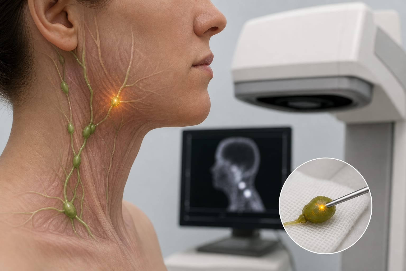
Painless left lateral neck mass present for over 5 years.
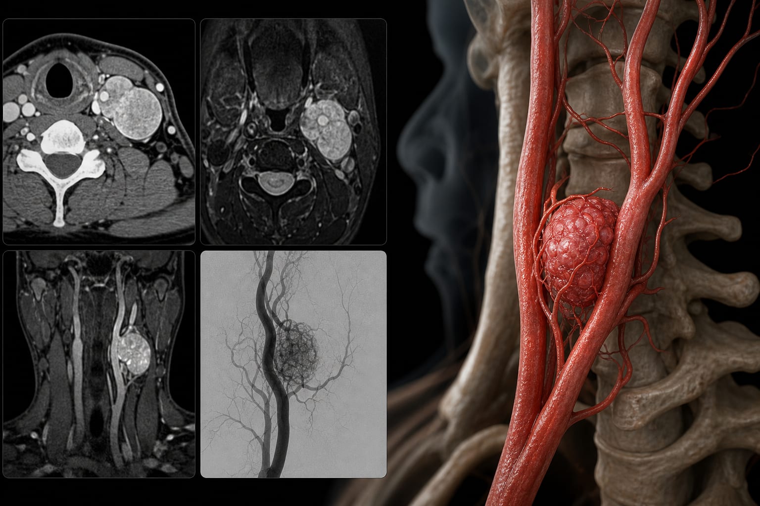
Diagnosis supported by ultrasonography, CTA, MRI, and DSA.
Surgical excision achieved with preservation of carotid arteries and nerves.
Histopathology confirmed diagnosis with a Ki-67 labeling index of approximately 5%.
Long-term follow-up showed no recurrence-related symptoms.
Guideline-Based Recommendations
Diagnosis
Utilize multimodal imaging including ultrasonography, CTA, MRI, and DSA for diagnosis.
Management
Surgical excision is recommended for resectable lesions.
Monitoring & Follow-up
Follow-up imaging and clinical assessment for recurrence.
Risks
Potential for vascular complications due to tumor's proximity to carotid arteries.
Patient & Prescribing Data
Young adults with carotid body paraganglioma.
Surgical excision can be performed with vessel preservation.
Clinical Best Practices
Employ a multimodal imaging approach for accurate diagnosis and surgical planning.
Ensure careful surgical technique to preserve surrounding vascular structures.