Functional ultrasound assessment of cerebral blood flow and brain connectivity in a pilocarpine-induced acute epileptic seizures in mice - Scorecard - MDSpire
Advertisement
Functional ultrasound assessment of cerebral blood flow and brain connectivity in a pilocarpine-induced acute epileptic seizures in mice
Clinical Scorecard: Evaluation of Cerebral Blood Flow and Brain Connectivity Using Functional Ultrasound in Mice with Pilocarpine-Induced Acute Seizures
At a Glance
Category
Detail
Condition
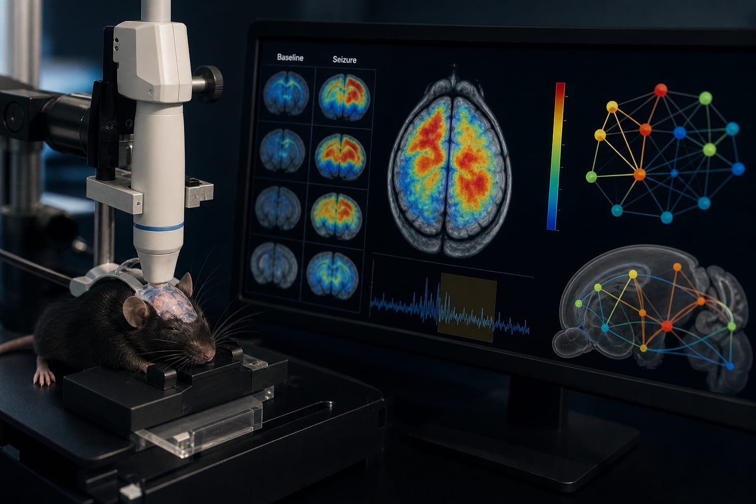
Temporal Lobe Epilepsy (TLE)
Key Mechanisms
Changes in cerebral blood volume (CBV) and functional connectivity during seizures.
Target Population
Mice with pilocarpine-induced acute seizures.
Care Setting
Animal research laboratory.
Key Highlights
Cerebral blood volume (CBV) changes were heterogeneous across the brain during acute seizures.
Most epilepsy-related brain regions showed an initial increase followed by a decrease in relative CBV (rCBV).
Functional connectivity of the brain underwent distinct modifications during acute seizure states.
Guideline-Based Recommendations
Diagnosis
Use functional ultrasound imaging (fUS) to assess changes in cerebral blood flow and connectivity.
Management
Monitor cerebral blood volume integrity as a potential mechanism for antiepileptic drug action.
Monitoring & Follow-up
Utilize fUS for real-time imaging of dynamic microvascular responses during seizures.
Risks
Cerebrovascular dysfunction can trigger and sustain seizures.
Patient & Prescribing Data
Not applicable; study conducted on mice.
Restoring CBV integrity is considered important in managing seizures.
Clinical Best Practices
Employ fUS for high-resolution imaging of brain blood flow changes during seizures.
Consider the role of vascular changes in the pathophysiology of temporal lobe epilepsy.