A Case of Fully Thrombosed Middle Cerebral Artery Aneurysm Resembling Cavernous Malformation: A Detailed Case Study with Surgical Video and Literature Review - Scorecard - MDSpire
Advertisement
A Case of Fully Thrombosed Middle Cerebral Artery Aneurysm Resembling Cavernous Malformation: A Detailed Case Study with Surgical Video and Literature Review
Clinical Scorecard: A Case of Fully Thrombosed Middle Cerebral Artery Aneurysm Resembling Cavernous Malformation: A Detailed Case Study with Surgical Video and Literature Review
At a Glance
Category
Detail
Condition
Key Mechanisms
Complete thrombosis due to thrombus and plaque leading to obliteration of the aneurysm lumen; consider adding physiological context.
Target Population
Care Setting
Key Highlights
Complete thrombosis of MCA aneurysms is rare and often misdiagnosed as cavernous malformations.
Initial imaging may show negative results for aneurysms, complicating diagnosis.
Surgical intervention can involve ultrasonic removal of thrombus and plaque with clip reconstruction.
Emphasize the importance of differential diagnosis to avoid mismanagement.
Guideline-Based Recommendations
Diagnosis
Consider differential diagnosis of aneurysmal thrombosis when encountering lesions in typical vascular locations, even with negative angiography.
Recommend pre-operative imaging techniques to enhance diagnostic accuracy.
Management
Surgical intervention is indicated for definitive treatment of completely thrombosed aneurysms.
Monitoring & Follow-up
Neuromonitoring during surgery is essential to ensure preservation of neurological function.
Risks
Misdiagnosis can lead to inappropriate management and potential complications.
Patient & Prescribing Data
Adults presenting with severe headaches and atypical imaging findings.
Postoperative outcomes can be favorable with appropriate surgical intervention.
Clinical Best Practices
Utilize high-resolution vessel wall MR imaging when available for better assessment of aneurysms.
Evaluate CT and MRI features carefully to differentiate between aneurysms and other lesions.
Involve multidisciplinary discussions for complex cases.
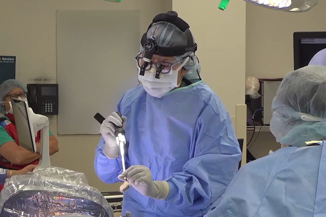
In this procedural case review, vascular surgeon Dr. Samuel Steerman and neurosurgeon Dr. Shannon Clark collaborate to perform an anterior lumbar interbody fusion (ALIF).