Clinical Scorecard: The 4 F Technique (Fat, Fascia, Fibrin, and Fat) for Reconstructing the Skull Base in Endoscopic Transorbital Surgery
At a Glance
Category
Detail
Condition
Osteodural defects following endoscopic transorbital skull base surgery
Key Mechanisms
Watertight closure of dural and bony defects using autologous fat grafts, fascia lata, and fibrin glue to prevent CSF leakage and related complications
Target Population
Adult patients (>18 years) undergoing superior eyelid endoscopic transorbital approach (SETOA) for intracranial lesions requiring dural opening or with CSF leak
Care Setting
Neurosurgical operating rooms performing endoscopic transorbital skull base surgery
Key Highlights
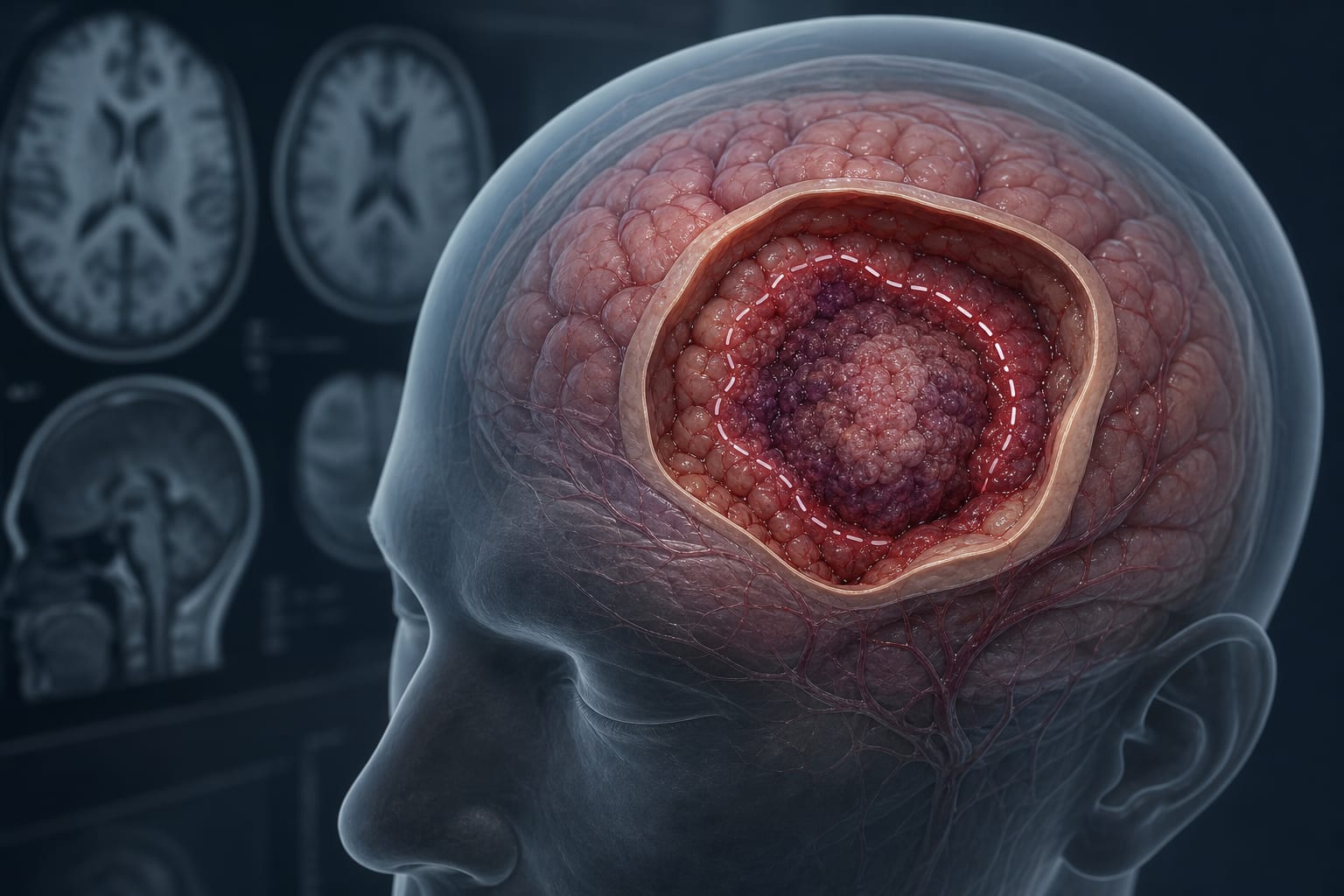
The 4F technique involves intradural autologous fat placement, extradurally applied fascia lata, fibrin glue fixation, and extradurally positioned autologous fat to fill dead space.
This method aims to restore barrier integrity between intra- and extradural compartments and eliminate dead space to reduce risks of CSF leak, meningitis, and pneumocephalus.
Applied in a series of 16 patients with various skull base lesions, the technique demonstrated effective reconstruction with attention to functional and esthetic outcomes.
Guideline-Based Recommendations
Diagnosis
Preoperative neurological and ophthalmological assessments to evaluate lesion impact.
Contrast-enhanced MRI to determine lesion location and growth pattern.
Identification of dural defects or CSF leaks intraoperatively.
Management
Perform superior eyelid endoscopic transorbital approach (SETOA) with or without extended variations depending on lesion extent.
Use the 4F reconstruction technique for osteodural defect repair: intradural fat graft, extradurally placed fascia lata, fibrin glue fixation, and extradurally placed fat graft.
Leave 2–3 mm of intentional proptosis post-reconstruction to accommodate postoperative edema resolution.
Monitoring & Follow-up
Postoperative assessment for CSF leak, enophthalmos, proptosis, diplopia, ocular paresis, and wound infection.
Follow-up ranging from 14 to 38 months to evaluate functional and esthetic outcomes.
Risks
Potential life-threatening complications include cerebrospinal fluid leakage, meningitis, and tension pneumocephalus if reconstruction is inadequate.
Risk of postoperative ocular complications such as diplopia or ocular paresis.
Possible wound infection related to surgical approach.
Patient & Prescribing Data
Adults undergoing SETOA for intracranial lesions with dural opening or CSF leak
The 4F technique provides a reliable, watertight reconstruction method using autologous tissues and fibrin glue, minimizing postoperative complications and supporting functional and esthetic recovery.
Clinical Best Practices
Ensure meticulous placement of autologous fat graft intradurally to seal dural defects.
Apply fascia lata extradurally to cover the osteodural defect securely.
Use fibrin glue to fix graft materials and enhance sealing.
Fill dead space extradurally with autologous fat to prevent postoperative complications.
Leave slight proptosis to accommodate postoperative edema and achieve binocular symmetry.
Perform careful suturing of periosteum, orbicularis oculi muscle, and eyelid skin to optimize healing.
by Sergio Corvino, Francesco Corrivetti, Giuseppe Catapano, Giuseppe Corazzelli, Antonio Colamaria, Edisher Maghalashvili, Iacopo Dallan, Domenico Di Maria, Germano Di Matteo, Giorgio Iaconetta, Matteo de Notaris