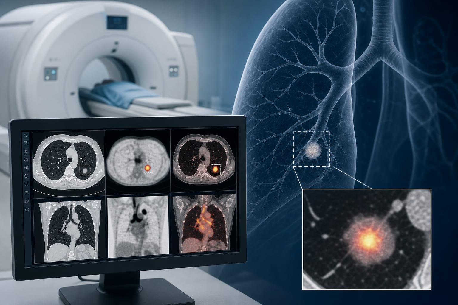
The role of 18F-FDG PET/CT in identifying risk factors for ground-glass nodules in invasive lung adenocarcinoma
-
By
-
Wenjun Bao
-
Chenmin Ding
-
Nan Li
-
Min Huang
-
Zaosheng Huang
-
Hailong Li
-
Jing Fan
-
Tiantian He
-
Xibao Mao
-
July 7, 2026
-
Clinical Scorecard: Utilizing 18F-FDG PET/CT to Assess Risk Factors Associated with Ground-Glass Nodules in Invasive Lung Adenocarcinoma
At a Glance
| Category | Detail |
|---|
| Condition | Invasive Lung Adenocarcinoma |
| Key Mechanisms | Utilization of 18F-FDG PET/CT to evaluate metabolic activity and morphological features of ground-glass nodules. |
| Target Population | Patients with surgically confirmed ground-glass nodule type lung adenocarcinoma. |
| Care Setting | Oncology and radiology departments for preoperative assessment. |
Key Highlights
- MPA and SPA subtypes of LUAD are associated with higher recurrence risks.
- SUVmax, nodule diameter, and lesion location are independent predictors of high-risk LUAD.
- The study developed a predictive model with AUCs of 0.934 (training) and 0.873 (test).
- PET/CT imaging can improve risk stratification for GGN-associated LUAD.
- Conventional FDG PET/CT parameters have limited diagnostic value for pure GGNs.
Guideline-Based Recommendations
Diagnosis
- Preoperative 18F-FDG PET/CT imaging is recommended for risk evaluation in LUAD.
Management
- Utilize PET/CT-derived metrics for stratifying risk in patients with GGN-associated LUAD.
Monitoring & Follow-up
- Regular follow-up imaging may be necessary for high-risk LUAD patients.
Risks
- Limited diagnostic value of FDG PET/CT for pure ground-glass nodules.
Patient & Prescribing Data
Patients with solitary ground-glass nodules and confirmed invasive adenocarcinoma.
Preoperative risk stratification can guide management decisions.
Clinical Best Practices
- Incorporate both PET metabolic indicators and CT morphological features for risk assessment.
- Ensure informed consent is obtained prior to imaging procedures.
Related Resources & Content