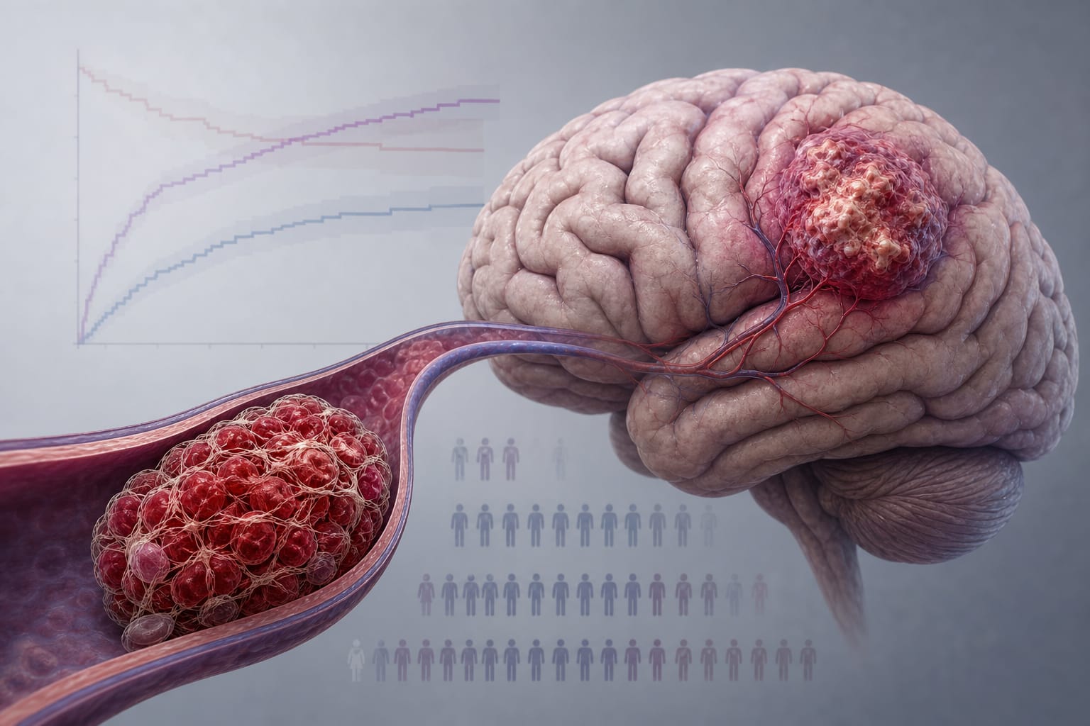
Blood neurofilament light chain measurements in adults with CNS histiocytic neoplasms
Clinical Scorecard: Measurement of Blood Neurofilament Light Chains in Adults Diagnosed with CNS Histiocytic Neoplasms
At a Glance
Category Detail
Condition CNS involvement in histiocytic neoplasms Key Mechanisms Accumulation of neoplastic histiocytic/dendritic cells causing CNS disease; neurofilament light chain (NfL) as a biomarker of axonal degeneration Target Population Adults diagnosed with histiocytic neoplasms including ECD, LCH, RDD with or without CNS involvement Care Setting Specialized neurology and hematology clinics with access to MRI and blood biomarker testing
Key Highlights
Elevated blood NfL levels are significantly associated with CNS involvement in histiocytic neoplasms. MRI with gadolinium enhancement remains the standard for detecting CNS disease and monitoring treatment response. NfL testing can be performed in blood using Simoa or Ella assays and correlates with CNS parenchymal involvement and enhancement. Guideline-Based Recommendations
Diagnosis
Use contrast-enhanced brain and spine MRI to evaluate CNS involvement in histiocytic neoplasms. Define CNS involvement by parenchymal T2 hyperintensity or gadolinium enhancement, meningeal or cranial nerve enhancement, or pituitary involvement. Consider blood NfL measurement as a biomarker to support CNS disease activity assessment. Management
Monitor CNS disease activity using serial MRI and blood NfL levels. Test for BRAF V600E and other relevant mutations to guide targeted therapies. Monitoring & Follow-up
Repeat MRI brain and spine with gadolinium to assess treatment response and disease progression. Serial blood NfL measurements can provide non-invasive monitoring of axonal injury and CNS disease activity. Risks
Comorbid neurologic, cardiac, and renal diseases may influence NfL levels and should be considered when interpreting results. Patient & Prescribing Data
Adults with histiocytic neoplasms, including those with CNS involvement
BRAF V600E mutation present in approximately half of patients with CNS involvement; mutation testing informs targeted treatment decisions.
Clinical Best Practices
Perform centralized pathological review to confirm histiocytic neoplasm subtype. Use standardized MRI protocols with gadolinium contrast for CNS disease evaluation. Incorporate blood NfL testing as an adjunct biomarker for CNS involvement and disease activity. Interpret NfL levels in context of patient age and comorbidities affecting NfL. Utilize multidisciplinary teams including neurology and hematology specialists for comprehensive care. References