Clinical Scorecard: Identifying Risk Factors and Prognostic Markers for Progressive Fibrosing Interstitial Lung Disease Using Deep Learning Techniques for CT Imaging Analysis
Increased fibrosis on CT scans, declining lung function (FVC), worsening symptoms leading to early mortality
Target Population
Patients with PF-ILD including idiopathic pulmonary fibrosis (IPF) and non-IPF ILD subtypes such as NSIP, CTD-ILD, and fibrotic hypersensitivity pneumonitis
Care Setting
Tertiary referral hospital with access to advanced imaging and pulmonary function testing
Key Highlights
PF-ILD is defined by relative decline in FVC and/or increased fibrosis on CT despite standard treatment.
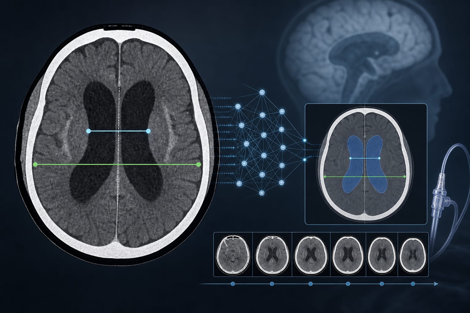
Visual CT assessment has significant inter- and intra-reader variability; quantitative CT (QCT) analysis improves evaluation accuracy.
Deep learning-based QCT techniques show promise in classifying ILD subtypes and predicting PF-ILD outcomes.
Guideline-Based Recommendations
Diagnosis
Use chest CT to identify ILD patterns (UIP, probable UIP, indeterminate/alternative) per recent IPF guidelines.
Apply visual criteria for fibrosis progression including traction bronchiectasis, ground-glass opacity with traction bronchiectasis, reticular opacity, honeycombing, and lung volume loss.
Incorporate quantitative CT analysis with deep learning-based texture segmentation for objective fibrosis and ILD extent assessment.
Management
Monitor forced vital capacity (FVC) decline over at least 24 months to define progression.
Consider tyrosine-kinase inhibitor therapy (e.g., nintedanib) for PF-ILD as per INBUILD study criteria.
Monitoring & Follow-up
Perform baseline and follow-up CT scans at intervals ≥24 months; intermediate 1-year CT scans may provide additional prognostic information.
Use quantitative CT metrics (fibrosis extent as sum of reticular opacity and honeycombing; total ILD extent including ground-glass opacity) to track disease progression.
Correlate CT findings with pulmonary function tests performed within 3 months of imaging.
Risks
Visual CT assessment variability may lead to inconsistent diagnosis and progression evaluation.
Early mortality risk is comparable between IPF and non-IPF PF-ILD patients with progressive fibrosis.
Exclusion of patients with acute exacerbations or significant pleural effusion is necessary for accurate imaging assessment.
Patient & Prescribing Data
Patients with PF-ILD including IPF and non-IPF ILD subtypes exhibiting progressive fibrosis
Nintedanib, a tyrosine-kinase inhibitor, has demonstrated efficacy in slowing progression in PF-ILD patients as defined by FVC decline and CT fibrosis progression.
Clinical Best Practices
Utilize deep learning-based quantitative CT analysis to reduce variability and improve accuracy in ILD subtype classification and fibrosis quantification.
Ensure CT imaging intervals of at least 24 months to reliably assess progression in PF-ILD.
Combine imaging findings with pulmonary function tests for comprehensive disease monitoring.
Engage experienced thoracic radiologists for visual CT pattern classification and fibrosis progression assessment.
Exclude confounding factors such as acute exacerbations, pleural effusions, and prior lung surgery when interpreting imaging for PF-ILD.