MR-guided microwave ablation of liver tumors: outcomes in local tumor control and determinants of treatment success
By
Vanessa F. Schmidt
Philipp Linden
Olaf Dietrich
Sinan Deniz
Daniel Puhr-Westerheide
Osman Öcal
Moritz L. Schnitzer
Florian Obereisenbuchner
Matthias Kassube
Mingming Wu
Luigi Nardone
Lars Grenacher
Florian Maier
Ricarda Seidensticker
Moritz Wildgruber
Jens Ricke
Matthias P. Fabritius
Max Seidensticker
July 4, 2026
Clinical Scorecard: Microwave Ablation of Hepatic Tumors Under MR Guidance: Efficacy in Local Control and Factors Influencing Treatment Success
At a Glance
Category Detail
Condition Hepatic Tumors Key Mechanisms Minimally invasive thermal ablation using microwave technology for local tumor control. Target Population Patients with primary and secondary liver malignancies. Care Setting Interventional radiology with MR guidance.
Key Highlights
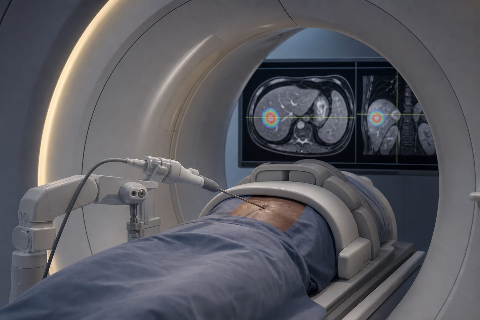
Microwave ablation (MWA) offers higher intratumoral temperatures and larger ablation volumes compared to radiofrequency ablation. MR imaging provides superior soft tissue contrast for accurate lesion localization and monitoring. Technical success rate of 95.8% was achieved with MR-guided MWA. Factors influencing treatment success include lesion size, type, and proximity to vascular structures. Complications were minimal, with only 2.5% experiencing postprocedural issues. Guideline-Based Recommendations
Diagnosis
Histopathological confirmation of liver malignancies is required prior to MWA. Management
Multidisciplinary tumor board involvement in treatment decision-making for MWA. Monitoring & Follow-up
MR thermometry should be utilized for continuous temperature monitoring during ablation. Risks
Potential complications include bleeding and pleural effusion, though these are rare. Patient & Prescribing Data
40 patients with a median age of 62.5 years, predominantly male.
Prior treatments were common, with 65% having received systemic therapy or other local ablative therapies.
Clinical Best Practices
Utilize MR imaging for precise targeting and monitoring during MWA. Ensure adequate safety margins from adjacent vascular structures during ablation. Conduct follow-up imaging to assess local tumor control post-ablation. Related Resources & Content