Comparison of the short-term efficacy of percutaneous endoscopic transforaminal lumbar interbody fusion with the application of a expandable versus PEEK cage for the treatment of lumbar degenerative disease - Scorecard - MDSpire
Conexiant’s news site is now MDSpire News. Learn more
Advertisement
Short-Term Outcomes of Percutaneous Endoscopic Transforaminal Lumbar Interbody Fusion Using Expandable Versus PEEK Cages for Lumbar Degenerative Disease
Clinical Scorecard: Short-Term Outcomes of Percutaneous Endoscopic Transforaminal Lumbar Interbody Fusion Using Expandable Versus PEEK Cages for Lumbar Degenerative Disease
At a Glance
Category
Detail
Condition
Lumbar disc herniation; eligibility criteria also included grade I–II degenerative or isthmic spondylolisthesis and segmental instability with disc herniation or stenosis.
Key Mechanisms
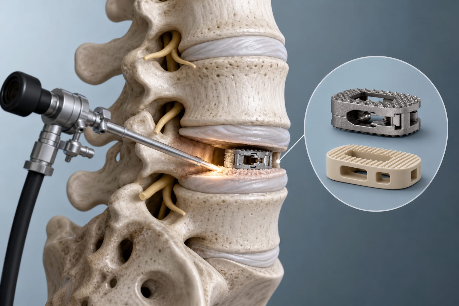
PE-TLIF involves endoscopic decompression, bone grafting, and interbody cage placement. Expandable cages are inserted at comparatively low volume and expanded in situ; static PEEK cages have fixed dimensions.
Target Population
Patients with lumbar disc herniation treated at one institution from May 2019 through February 2021; eligibility criteria included concordant imaging and symptoms persisting despite three months of regular, systematic conservative treatment, with specified additional diagnoses and exclusions.
Care Setting
Surgical treatment of selected lumbar degenerative disease using percutaneous endoscopic transforaminal lumbar interbody fusion.
Key Highlights
This retrospective investigation compared clinical and radiological outcomes of expandable cages and static PEEK cages during PE-TLIF.
The study enrolled patients treated at one institution between May 2019 and February 2021; all procedures were performed by one senior spinal surgeon.
Eligibility required symptoms persisting despite three months of regular, systematic conservative treatment and imaging concordant with clinical findings.
Exclusions included severe lumbar spinal stenosis, severe osteoporosis, grade III–IV spondylolisthesis, and prior lumbar surgery.
The supplied article excerpt does not report comparative outcome results.
Guideline-Based Recommendations
Diagnosis
Eligibility criteria included lumbar symptoms with imaging findings concordant with clinical symptoms and signs.
Specified conditions included lumbar disc herniation, grade I–II degenerative or isthmic spondylolisthesis, and segmental instability with disc herniation or stenosis.
Management
The article states that surgery was generally recommended for intractable symptoms despite conservative treatment or in the presence of cauda equina syndrome.
The described PE-TLIF procedure included endoscopic decompression, nucleus pulposus removal, bone grafting, and interbody cage placement.
Monitoring & Follow-up
Risks
The introduction identifies potential risks of static PEEK cages, including postoperative migration and subsidence.
The article describes potential risks of larger-device implantation, including nerve root or dural sac injury associated with a larger working channel and additional bone removal.
Open lumbar fusion procedures are described as carrying risks including blood loss, paraspinal soft-tissue injury, chronic low back pain, dural tear, wound infection or poor healing, and nerve root injury.
Patient & Prescribing Data
Patients with lumbar disc herniation treated at the institution from May 2019 through February 2021 were retrospectively enrolled. Eligibility required concordant imaging and symptoms persisting despite three months of regular, systematic conservative treatment; additional eligibility criteria and exclusions are described in the article.
The study compared expandable and static PEEK cages in PE-TLIF. The supplied excerpt describes the rationale and eligibility criteria but provides no comparative results or prescribing conclusions.
Clinical Best Practices
The study required symptoms to persist despite three months of regular, systematic conservative treatment for eligibility.
The described procedure used fluoroscopic identification of the target level and an endoscopic working channel for decompression and interbody fusion.
The article describes preparing bone removed during decompression for grafting.