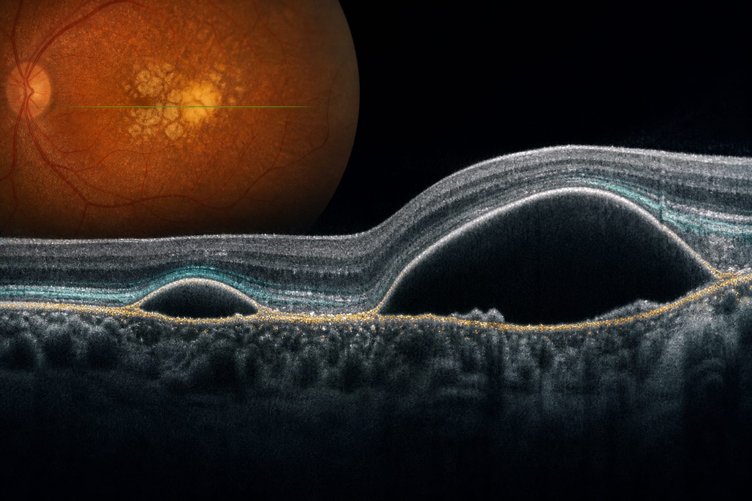
Elevations of the Retinal Pigment Epithelium in Age-Related Macular Degeneration: Shallow and Bullous Forms
-
By
-
Jost B. Jonas
-
Songhomitra Panda-Jonas
-
Jie Xue
-
Rahul A. Jonas
-
Ya Xing Wang
-
September 25, 2026
Clinical Scorecard: Elevations of the Retinal Pigment Epithelium in Age-Related Macular Degeneration: Shallow and Bullous Forms
At a Glance
| Category | Detail |
|---|
| Condition | Age-Related Macular Degeneration (AMD) |
| Key Mechanisms | Separation of the retinal pigment epithelium (RPE) from Bruch's membrane, leading to elevations and detachments. |
| Target Population | Individuals aged 50 years and older with early-stage or intermediate-stage AMD. |
| Care Setting | Population-based cohort study |
Key Highlights
- RPE elevations and detachments are part of the morphologic spectrum of AMD.
- Shallow and bullous RPE elevations are associated with a higher risk of AMD progression.
- The study included a large cohort with participants without maculopathies.
Guideline-Based Recommendations
Diagnosis
- AMD diagnosed based on criteria from the Beckman Initiative for Macular Research Classification Committee.
Management
- Assessment of RPE elevations and detachments using optical coherence tomography (OCT).
Monitoring & Follow-up
- Longitudinal studies to evaluate the progression of AMD in relation to RPE changes.
Risks
- Higher risk of progression to geographic atrophy associated with RPE elevations and detachments.
Patient & Prescribing Data
Participants aged 50 years and older with early-stage or intermediate-stage AMD.
No specific treatment insights provided; focus on prevalence and associations.
Clinical Best Practices
- Utilize OCT for detailed assessment of RPE elevations and detachments.
- Consider longitudinal monitoring of patients with identified RPE changes.
Related Resources & Content