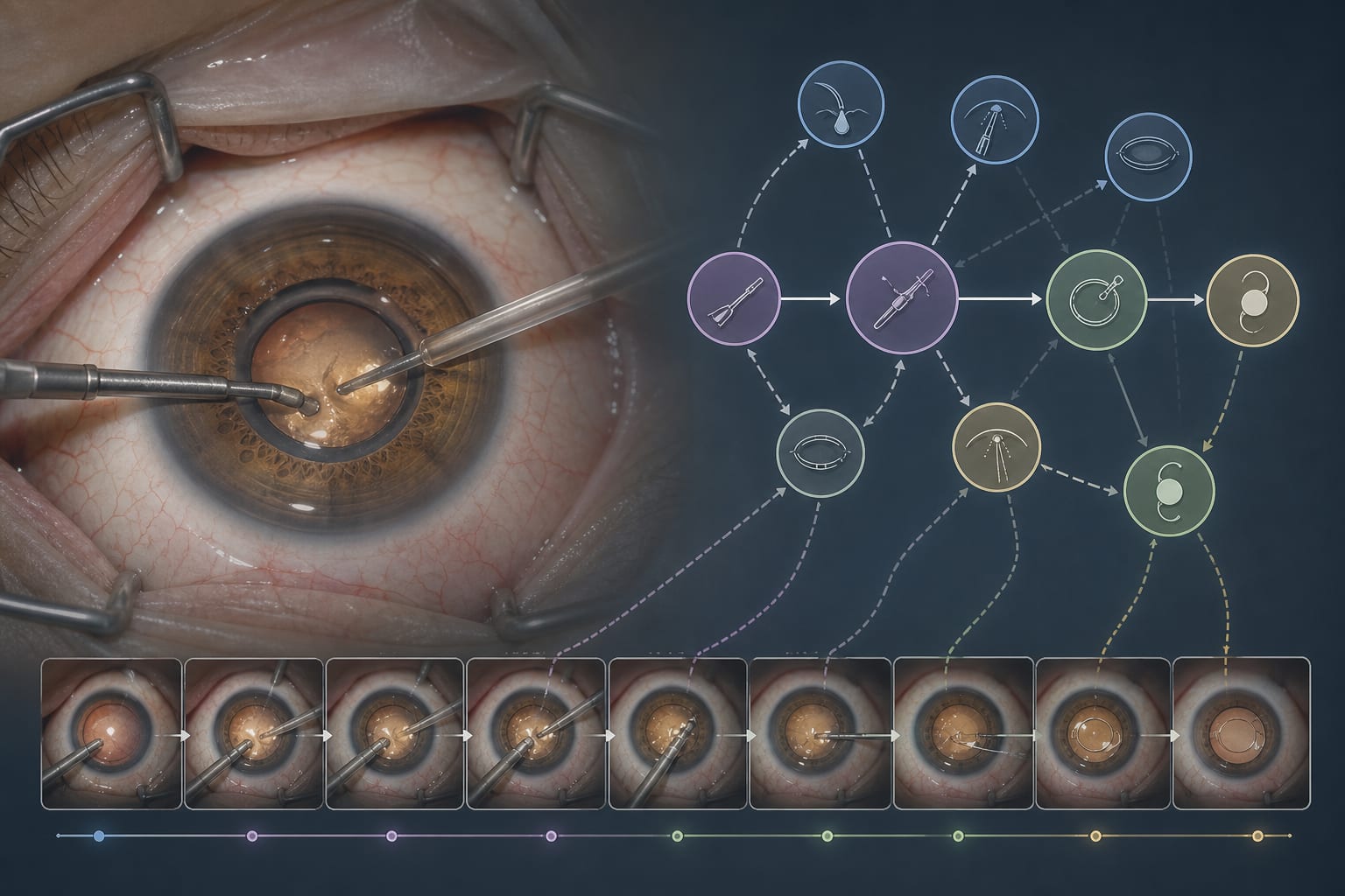
Removal of Implantable Collamer Lenses Due to Elevated Intraocular Pressure in a Patient with Severe Hyperopia: A Case Study
-
By
-
Liuqing Cui
-
Karl Mercieca
-
Leonie Bourauel
-
March 24, 2026
Clinical Scorecard: Removal of Implantable Collamer Lenses Due to Elevated Intraocular Pressure in a Patient with Severe Hyperopia: A Case Study
At a Glance
| Category | Detail |
|---|---|
| Condition | Elevated intraocular pressure (IOP) and angle closure following Implantable Collamer Lens (ICL) implantation in severe hyperopia |
| Key Mechanisms | ICLs without central aperture causing angle closure, peripheral anterior synechiae, iris adhesions leading to IOP elevation and risk of secondary glaucoma |
| Target Population | Patients with high hyperopia and astigmatism who have undergone ICL implantation |
| Care Setting | Ophthalmology clinics with surgical and medical glaucoma management capabilities |
Key Highlights
- ICL implantation without central port can lead to long-term complications including elevated IOP and secondary angle closure glaucoma.
- Medical therapy with multiple IOP-lowering agents may be insufficient to control IOP in some patients after ICL implantation.
- Combined explantation of ICL and cataract surgery with posterior chamber IOL implantation can effectively reduce IOP and inflammation.
Guideline-Based Recommendations
Diagnosis
- Perform slit-lamp examination to assess ICL position and iris adhesions.
- Use gonioscopy to evaluate chamber angle status and presence of peripheral anterior synechiae.
- Conduct visual field testing and OCT to assess for glaucomatous damage.
Management
- Initiate IOP-lowering medications including prostaglandin analogs, carbonic anhydrase inhibitors, and alpha-agonists.
- Consider combined surgical explantation of ICL and phacoemulsification with posterior chamber IOL implantation in cases of inadequate IOP control or angle closure.
- Administer perioperative anti-inflammatory treatment including topical steroids and subconjunctival dexamethasone.
Monitoring & Follow-up
- Regular follow-up every 3-4 months to monitor IOP and anterior chamber inflammation.
- Assess visual acuity and anterior chamber reaction postoperatively.
- Monitor for postoperative complications such as fibrin deposition and iris defects.
Risks
- Risk of iris trauma and prolapse during ICL explantation due to adhesions.
- Potential postoperative inflammation requiring intensive steroid therapy.
- Loss of accommodation following cataract surgery with posterior chamber IOL implantation.
Patient & Prescribing Data
38-year-old woman with high hyperopia and family history of glaucoma post-ICL implantation
Multiple IOP-lowering agents (bimatoprost, dorzolamide, clonidine) were insufficient; surgical explantation combined with cataract surgery improved IOP control and visual acuity.
Clinical Best Practices
- Careful preoperative assessment of angle status and iris adhesions before ICL explantation.
- Use of iris retractors and intravenous mannitol to minimize intraoperative iris trauma.
- Aggressive postoperative anti-inflammatory therapy including topical steroids and subconjunctival injections to control inflammation.
- Patient counseling regarding risks of surgery, loss of accommodation, and potential postoperative visual symptoms such as glare.
Related Resources & Content
- Reference 1 - Safety and efficacy of PC pIOL implantation in high hyperopia
- Reference 2 - Complications associated with ICLs without central aperture
Based on findings from:
Removal of Implantable Collamer Lenses Due to Elevated Intraocular Pressure in a Patient with Severe Hyperopia: A Case Study
Liuqing Cui, Karl Mercieca, Leonie Bourauel. Bmc Ophthalmology, 2026.
https://link.springer.com/article/10.1186/s12886-026-04757-1
This content is an AI-generated, fully rewritten summary based on a published scholarly article. It does not reproduce the original text and is not a substitute for the original publication. Readers are encouraged to consult the source for full context, data, and methodology.