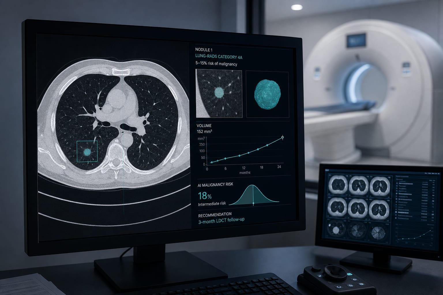
Clinical Scorecard: Validation of CT Radiomics Models for Predicting Local Tumor Progression in Colorectal Liver Metastases Following Ablation
At a Glance
Category
Detail
Condition
Colorectal liver metastases (CRLM) treated with thermal ablation
Key Mechanisms
Prediction of local tumor progression (LTP) using radiomics features from post-ablation contrast-enhanced CT images combined with clinical parameters
Target Population
Patients with CRLM undergoing thermal ablation (microwave or radiofrequency ablation)
Care Setting
Multicentre tertiary care institutions with imaging and interventional radiology capabilities
Key Highlights
Thermal ablation is an alternative to resection for CRLM but has LTP rates of 6–46%.
LTP detection by contrast-enhanced CT has limited sensitivity (53%) due to similar imaging characteristics of post-ablation effects and recurrence.
Combined clinical-radiomics models from post-ablation CT images can predict LTP with good performance (c-statistic 0.78), enabling risk stratification.
Guideline-Based Recommendations
Diagnosis
Use contrast-enhanced portal venous phase CT 2–8 weeks post-ablation for initial assessment.
Define LTP as new tumor foci within 10 mm of the ablation zone on follow-up imaging within 24 months.
Employ MRI or PET-CT as problem-solving modalities when CT findings are equivocal.
Management
Consider complementary treatment promptly for patients predicted to have high risk of LTP based on radiomics models.
De-intensify follow-up imaging schedules for patients at low risk of LTP to reduce unnecessary imaging burden.
Monitoring & Follow-up
Schedule follow-up contrast-enhanced CT every 3 months in the first year post-ablation, then every 6 months up to 5 years.
Monitor for new lesions within 10 mm of the ablation zone to detect LTP early.
Risks
Incomplete ablation (residual disease, margins <5 mm) increases risk of LTP and should be excluded from predictive modeling.
Post-ablation imaging artifacts (e.g., air, abscess) can complicate LTP detection.
Patient & Prescribing Data
Patients with histopathologically confirmed CRLM treated with thermal ablation and available portal venous phase CT imaging post-ablation
Radiomics combined with clinical data can stratify patients by LTP risk, guiding personalized follow-up and timely additional interventions.
Clinical Best Practices
Ensure high-quality contrast-enhanced CT imaging with standardized acquisition protocols for reliable radiomics analysis.
Exclude patients with >5 ablated CRLM, lesions >3 cm, diffuse liver disease, or prior liver treatments affecting parenchyma to maintain model validity.
Perform manual delineation of ablation zone and peri-ablational rim for radiomics feature extraction.
Use external validation cohorts to confirm generalizability of predictive models before clinical implementation.
by Denise J. van der Reijd, Corentin Guerendel, Femke C. R. Staal, Milou P. Busard, Mateus De Oliveira Taveira, Elisabeth G. Klompenhouwer, Koert F. D. Kuhlmann, Adriaan Moelker, Cornelis Verhoef, Martijn P. A. Starmans, Doenja M. J. Lambregts, Regina G. H. Beets-Tan, Sean Benson, Monique Maas