A visual mining analysis of middle meningeal embolization and other factors associated with recurrence requiring re-operation in subdural hematomas: a single-center series - Scorecard - MDSpire
Advertisement
A visual mining analysis of middle meningeal embolization and other factors associated with recurrence requiring re-operation in subdural hematomas: a single-center series
Clinical Scorecard: Analysis of Visual Data on Middle Meningeal Embolization and Factors Linked to Recurrence Necessitating Reoperation in Subdural Hematomas: Insights from a Single-Center Study
At a Glance
Category
Detail
Condition
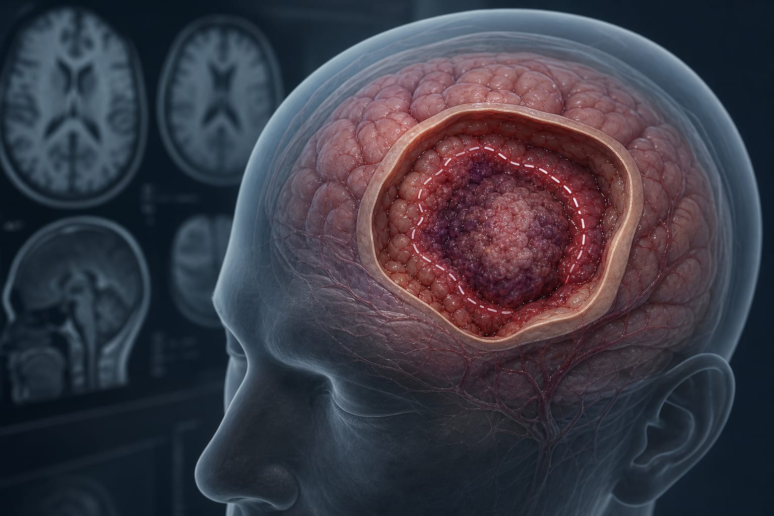
Chronic subdural hematoma (cSDH)
Key Mechanisms
Recurrence linked to hematoma internal architecture, volume, postoperative residual volume, pneumocephalus, brain atrophy, coagulation profile, and antiplatelet therapy; middle meningeal artery embolization (MMAE) as adjunct or stand-alone treatment
Target Population
Predominantly individuals over 65 years with cSDH
Care Setting
Neurosurgical care including surgical evacuation and adjunctive neurointerventional procedures
Key Highlights
cSDH recurrence rates post-surgery can reach up to 50%, independent of surgical technique.
Risk factors for recurrence include Nakaguchi hematoma type, pre/postoperative hematoma volumes, pneumocephalus, brain atrophy, bilateral hematomas, platelet count, and antiplatelet therapy.
MMA embolization is an emerging adjunctive treatment to reduce recurrence, performed via selective catheterization and PVA particle injection.
Guideline-Based Recommendations
Diagnosis
Use non-contrast brain CT scans with volumetric assessment for diagnosis and monitoring.
Classify hematoma internal architecture using Nakaguchi classification to assess recurrence risk.
Management
Standard surgical evacuation via burr hole or minicraniotomy with subdural drain placement.
Consider adjunctive MMA embolization starting October 2022 to reduce recurrence risk.
Manage antiplatelet and anticoagulation therapy carefully during perioperative period.
Monitoring & Follow-up
Postoperative CT within 24 hours to assess residual hematoma volume and pneumocephalus.
Follow-up imaging to monitor hematoma volume and neurological status.
Monitor coagulation parameters including platelet count, aPTT, and INR.
Risks
High recurrence risk with separated Nakaguchi hematoma type and large pre/postoperative volumes.
Increased recurrence associated with postoperative pneumocephalus, brain atrophy, bilateral hematomas.
Patients with chronic subdural hematoma undergoing surgical evacuation, predominantly elderly
Adjunctive MMA embolization introduced as standard care since October 2022; antiplatelet and anticoagulation therapies require careful management to mitigate recurrence risk.
Clinical Best Practices
Perform MMA embolization under local anesthesia or conscious sedation using standardized technique by experienced neurointerventionalists.
Use volumetric CT measurements pre- and postoperatively to stratify recurrence risk and guide management.
Apply Nakaguchi classification combined with volumetric parameters for risk assessment.
Maintain subdural drain placement post-surgery and remove within 24 hours.
Monitor and adjust antiplatelet and anticoagulation therapy perioperatively to balance bleeding and recurrence risks.