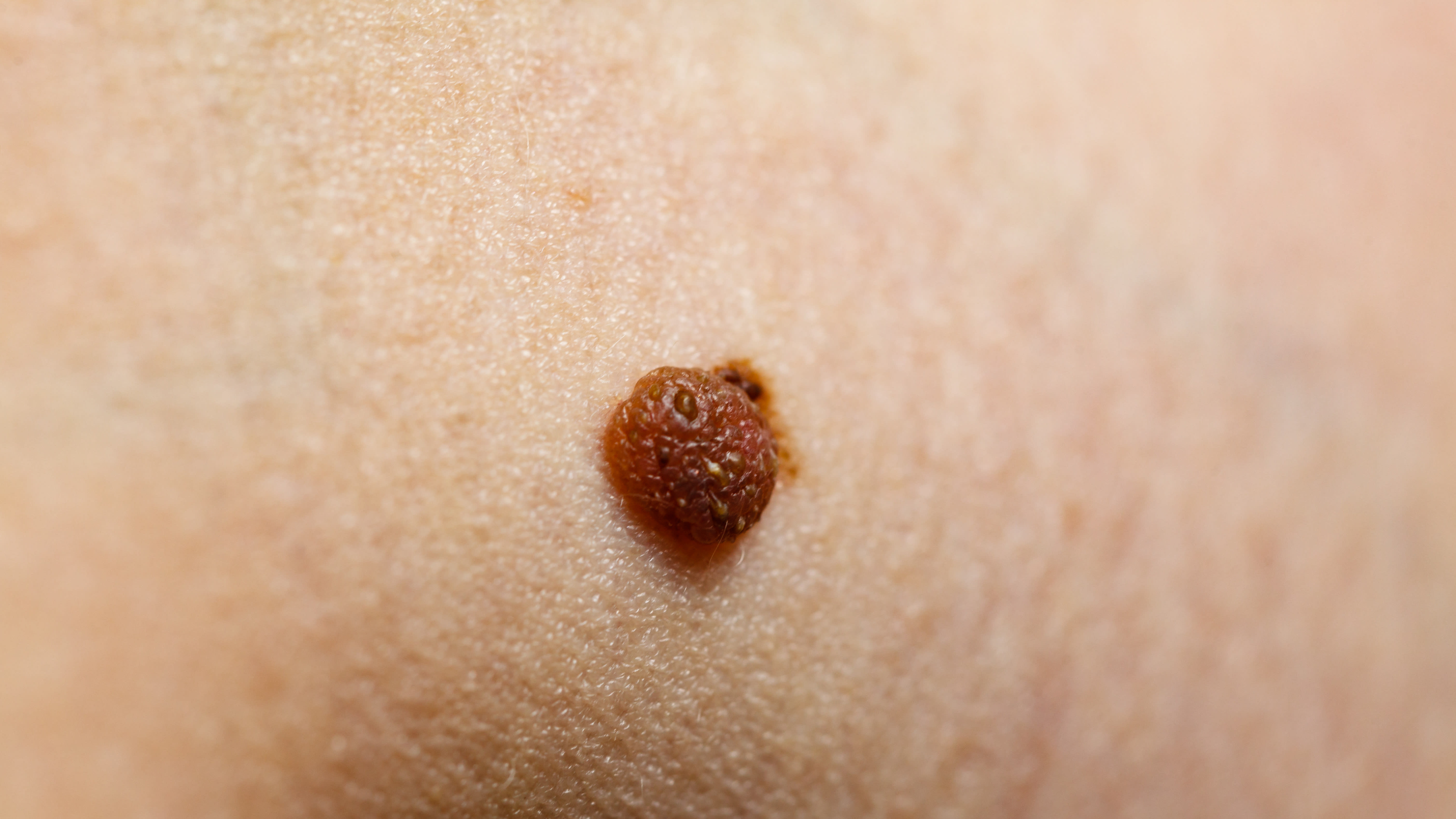
Robust Framework Utilizing Deep Neural Networks for Automated Segmentation and Analysis of Skin Lesions
Clinical Scorecard: Robust Framework Utilizing Deep Neural Networks for Automated Segmentation and Analysis of Skin Lesions
At a Glance
Category Detail
Condition Skin cancers including melanoma and non-melanoma skin cancers Key Mechanisms Automated segmentation of dermoscopic images using deep neural networks combined with traditional image enhancement techniques Target Population Patients with skin lesions suspected of melanoma or non-melanoma skin cancers Care Setting Dermatology clinics and computer-aided diagnosis (CAD) systems in clinical settings
Key Highlights
Melanoma is an aggressive skin cancer with high mortality requiring early detection and intervention. Visual inspection by dermatologists is subjective; automated segmentation improves diagnostic consistency and accuracy. The proposed hybrid framework combines morphological operations, Wiener filtering, and deep neural networks for robust lesion segmentation. Guideline-Based Recommendations
Diagnosis
Utilize dermoscopy for enhanced visualization of skin lesions. Incorporate automated segmentation techniques to reduce subjectivity in lesion boundary delineation. Apply pre-processing methods to improve image quality before segmentation. Management
Early detection and timely treatment of melanoma are critical to improve survival rates. Use computer-aided diagnosis systems integrating deep learning for therapeutic planning. Monitoring & Follow-up
Regular follow-up with dermoscopic imaging and automated analysis to track lesion changes. Monitor segmentation accuracy and update algorithms with diverse datasets for generalization. Risks
Subjectivity in visual inspection may lead to inconsistent diagnoses. Artifacts such as hair, blood vessels, and markings can interfere with image analysis. High computational demands and limited generalization of some advanced AI models. Patient & Prescribing Data
Individuals undergoing evaluation for suspicious skin lesions
Automated segmentation supports accurate diagnosis and informs appropriate treatment decisions, potentially reducing invasive procedures for non-melanoma cancers.
Clinical Best Practices
Combine traditional image enhancement with deep learning for improved segmentation accuracy. Employ U-Net and its variants for effective medical image segmentation with attention to model efficiency. Address image artifacts and lesion variability through robust pre-processing and post-processing techniques. Validate segmentation algorithms on diverse and complex datasets such as ISIC 2017. References