Intra-procedural CT control versus ultrasound only in ultrasound-guided thermal ablation of colorectal liver metastases: a single-centre cohort study - Scorecard - MDSpire

Intra-procedural CT control versus ultrasound only in ultrasound-guided thermal ablation of colorectal liver metastases: a single-centre cohort study

  • By

  • Trygve Syversveen

  • Ingrid Schrøder Hansen

  • Knut Brabrand

  • Kristoffer Watten Brudvik

  • Ida Björk

  • Daniel Østergaard

  • Olaug Villanger

  • Bård Ingvald Røsok

  • Kristoffer Lassen

  • Bjørn von Gohren Edwin

  • Sheraz Yaqub

  • Åsmund Avdem Fretland

  • May 22, 2026

  • 0 min

Share

Clinical Scorecard: Comparative Analysis of Intra-procedural CT Guidance versus Ultrasound Alone in Thermal Ablation for Colorectal Liver Metastases: A Single-Center Cohort Investigation

At a Glance

CategoryDetail
Condition
Key MechanismsThermal ablation using ultrasound (US) and computed tomography (CT) guidance, emphasizing the comparative effectiveness.
Target Population
Care Setting

Key Highlights

  • Local tumor progression (LTP) rates after ablation vary from 12 to 54%, influenced by imaging guidance.
  • CT guidance improves margin confirmation compared to visual assessment, particularly in challenging cases.

Guideline-Based Recommendations

Diagnosis

    Management

      Monitoring & Follow-up

      • P
      • o
      • s
      • t
      • -
      • p
      • r
      • o
      • c
      • e
      • d
      • u
      • r
      • a
      • l
      • s
      • u
      • r
      • v
      • e
      • i
      • l
      • l
      • a
      • n
      • c
      • e
      • i
      • n
      • c
      • l
      • u
      • d
      • e
      • s
      • c
      • o
      • n
      • t
      • r
      • a
      • s
      • t
      • -
      • e
      • n
      • h
      • a
      • n
      • c
      • e
      • d
      • C
      • T
      • e
      • v
      • e
      • r
      • y
      • 6
      • m
      • o
      • n
      • t
      • h
      • s
      • ,
      • w
      • i
      • t
      • h
      • s
      • p
      • e
      • c
      • i
      • f
      • i
      • c
      • a
      • t
      • t
      • e
      • n
      • t
      • i
      • o
      • n
      • t
      • o
      • L
      • T
      • P
      • r
      • a
      • t
      • e
      • s
      • .

      Risks

      • I
      • n
      • s
      • u
      • f
      • f
      • i
      • c
      • i
      • e
      • n
      • t
      • m
      • a
      • r
      • g
      • i
      • n
      • s
      • m
      • a
      • y
      • l
      • e
      • a
      • d
      • t
      • o
      • l
      • o
      • c
      • a
      • l
      • t
      • u
      • m
      • o
      • r
      • p
      • r
      • o
      • g
      • r
      • e
      • s
      • s
      • i
      • o
      • n
      • ;
      • e
      • m
      • p
      • h
      • a
      • s
      • i
      • z
      • e
      • t
      • h
      • e
      • i
      • m
      • p
      • o
      • r
      • t
      • a
      • n
      • c
      • e
      • o
      • f
      • a
      • d
      • e
      • q
      • u
      • a
      • t
      • e
      • m
      • a
      • r
      • g
      • i
      • n
      • a
      • s
      • s
      • e
      • s
      • s
      • m
      • e
      • n
      • t
      • .

      Patient & Prescribing Data

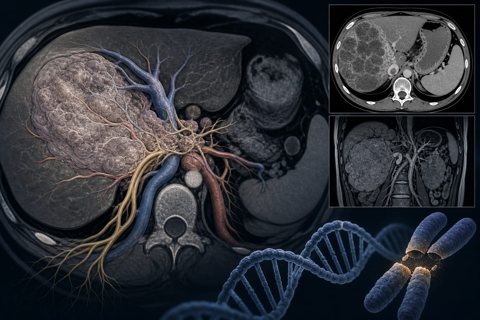
      Ablation chosen for small or deeply located tumors, frail patients, or technically challenging resections, with specific criteria outlined.

      Clinical Best Practices

      • Aim for a minimum ablation margin of 5 mm, ideally 10 mm, supported by recent studies.
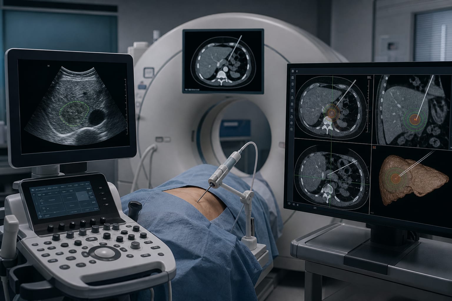
      • Use image fusion with recent CT or MRI for enhanced lesion localization, as per current best practices.
      • Perform CEUS control to assess the ablation zone post-treatment, referencing relevant clinical guidelines.

      Related Resources & Content

      Original Source(s)

      Related Content