Late enhancement and washout maps for distinguishing tumor progression from radiation necrosis in malignant brain tumors: an exploratory study - Scorecard - MDSpire
Clinical Scorecard: An Exploratory Study of Late Enhancement and Washout Maps to Differentiate Tumor Progression from Radiation Necrosis in Malignant Brain Tumors
At a Glance
Category
Detail
Condition
Suspected tumor progression, radiation necrosis, or pseudoprogression in adults with malignant brain tumors after treatment.
Key Mechanisms
Co-register post-contrast T1-weighted images acquired 5–9 and 25–29 minutes after contrast administration, normalize intensity, and subtract voxel-wise; maps display washout in red and delayed enhancement in green.
Target Population
Adults with histologically confirmed glioblastoma, other WHO grade 3–4 glioma, CNS lymphoma, or brain metastasis whose follow-up MRI after surgery and radiochemotherapy showed a newly appearing contrast-enhancing lesion.
Care Setting
Single-center retrospective observational imaging study using follow-up MRI.
Key Highlights
The study examined whether delayed-enhancement and washout maps could help distinguish tumor progression from radiation necrosis.
Post-contrast T1-weighted images were acquired at 5–9 minutes and again at 25–29 minutes.
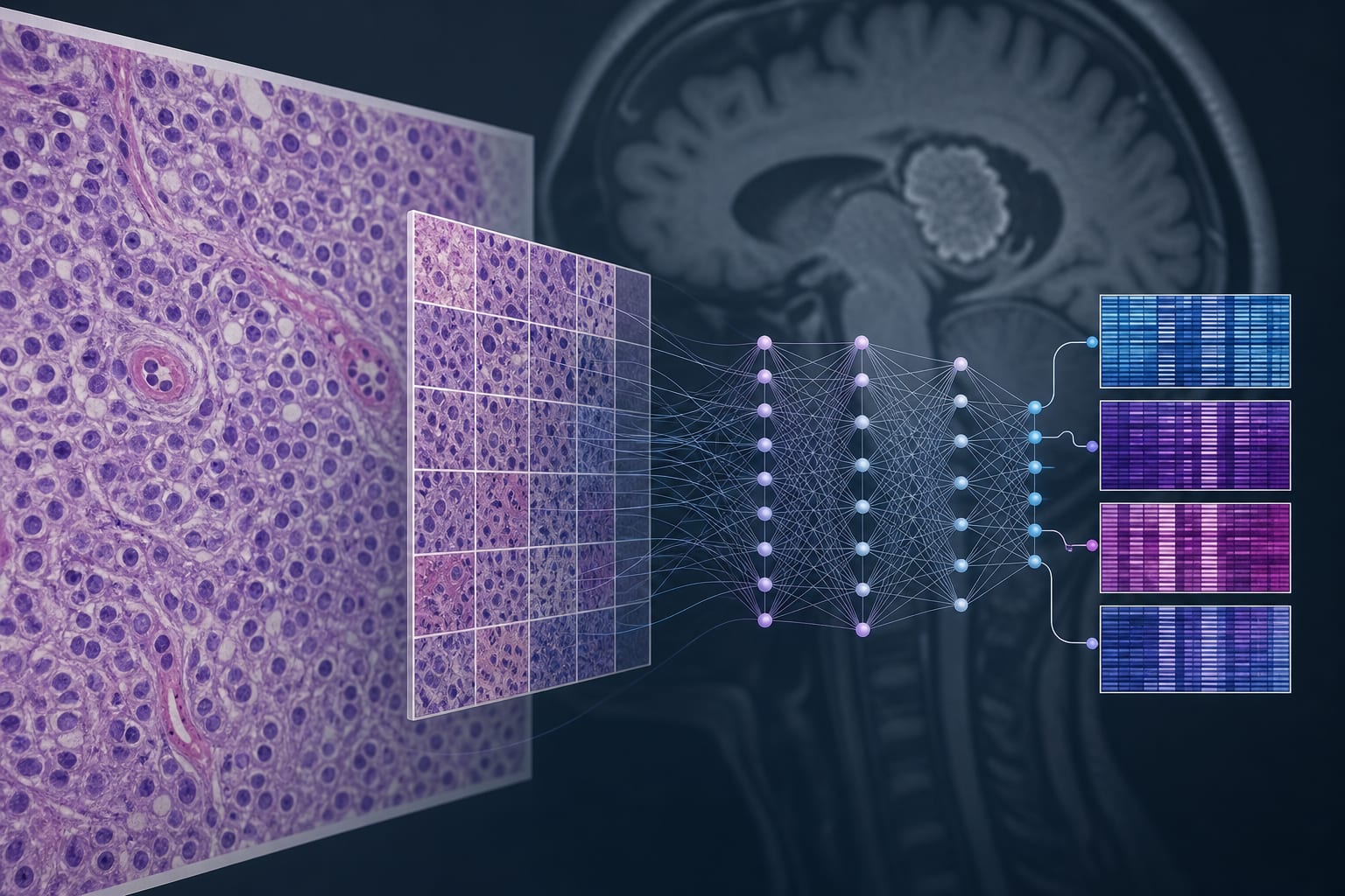
Washout maps were generated by co-registration, intensity normalization, and voxel-wise subtraction of the two T1-weighted datasets.
The MRI protocol also included DSC perfusion MRI and diffusion-weighted imaging.
Guideline-Based Recommendations
Diagnosis
The article describes an exploratory imaging method, not a clinical guideline or validated diagnostic recommendation.
Eligible cases had newly appearing contrast-enhancing lesions on follow-up MRI after surgery and radiochemotherapy.
Management
The source does not provide treatment recommendations based on washout-map findings.
Monitoring & Follow-up
The source excerpt does not specify a monitoring schedule or reference-standard follow-up procedure.
Risks
The introduction notes that radiation necrosis and pseudoprogression may resemble tumor progression on conventional contrast-enhanced MRI.
Motion artifacts, absent contrast enhancement, or insufficient measurable enhancing volume can prevent washout-map computation.
Patient & Prescribing Data
Adults aged 18 years or older with histologically confirmed glioblastoma, other WHO grade 3–4 glioma, CNS lymphoma, or brain metastasis and newly appearing enhancing lesions on follow-up MRI after surgery and radiochemotherapy.
The study evaluated imaging after surgery and radiochemotherapy. It reports no medication or treatment regimen recommendations.
Clinical Best Practices
The introduction describes difficulty distinguishing treatment-related changes from progression using conventional contrast-enhanced MRI.
The study protocol used paired post-contrast T1-weighted acquisitions at 5–9 and 25–29 minutes.
The study also assessed DSC perfusion and diffusion-weighted imaging alongside washout maps.
The introduction states that amino acid PET, particularly FET PET, may aid distinction when MRI findings are inconclusive; availability varies.
by Eya Khadhraoui, Leon Schmidt, Stefan Klebingat, I Erol Sandalcioglu, Roland Schwab, Philipp Arndt, Daniel Behme, Klaus-Peter Stein, Sebastian Johannes Müller
Experts across Baptist Health Herbert Wertheim Cancer Institute and Eugene M. & Christine E. Lynn Cancer Institute will present new radiation oncology research and clinical expertise at the 2026 Annual Meeting of the American Society for Radiation Oncology (ASTRO), September 26-30, 2026, in Boston.