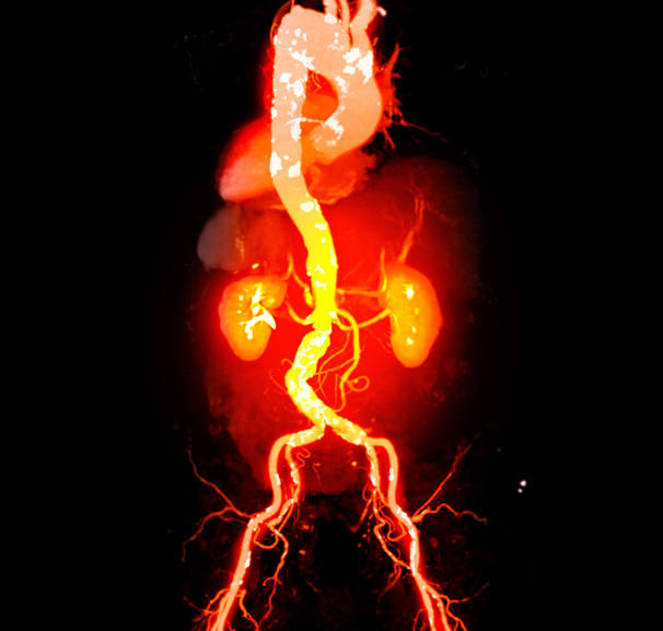
Lumbar disc herniation may mask coral reef aorta
-
By
-
Andrea Surnit
-
October 16, 2026
-
4 min
Clinical Scorecard: Lumbar disc herniation may mask coral reef aorta
At a Glance
| Category | Detail |
|---|---|
| Condition | Coral reef aorta causing near-occlusion of the infrarenal abdominal aorta and bilateral lower extremity ischemia |
| Key Mechanisms | Progressive protruding calcified aortic plaques narrowed the aortic lumen; coexisting lumbar disc herniation may have obscured recognition of vascular disease. |
| Target Population | Patients with unexplained, progressive bilateral lower extremity ischemic symptoms, including those with coexisting conditions that may mask vascular disease |
| Care Setting | Clinical evaluation and vascular imaging, with individualized consideration of open surgical or endovascular treatment |
Key Highlights
- A 50-year-old man with lumbar disc herniation developed progressive bilateral leg symptoms, intermittent claudication, and rest pain.
- Serial CT over 6 years showed progressive calcification extending from the posterior aortic wall into the lumen, causing near-occlusion.
- Open aortic graft replacement was selected because extensive circumferential calcification raised concerns about distal embolization and inadequate stent expansion.
- After surgery, symptoms resolved and ABI improved; at 1 year, the patient remained asymptomatic and CT showed graft and inferior mesenteric artery patency.
- The single-patient report cannot establish comparative treatment effectiveness or longer-term graft durability.
Guideline-Based Recommendations
Diagnosis
- Consider coral reef aorta in patients with unexplained lower extremity ischemia, particularly when symptoms are bilateral, progressive, or disproportionate to peripheral arterial findings.
- Clinical assessment in this case included physical examination, ABI measurements, and abdominal CT.
- Serial CT can characterize the extent and progression of aortic calcification.
Management
- Individualize treatment selection according to anatomical characteristics and patient risk profile.
- Endovascular treatment may be considered in selected patients with high surgical risk or limited disease extent.
- Extensive calcification may restrict stent expansion and increase distal embolization or vessel injury risk; open reconstruction was used in this advanced case.
Monitoring & Follow-up
- Postoperative assessment included ABI, symptom status, lower extremity pulses, and graft patency.
- At 1-year follow-up, ABI and contrast-enhanced CT were used to assess sustained improvement and graft patency.
Risks
- Extensive circumferential calcification may increase the risk of distal embolization, inadequate stent expansion, or vessel injury during endovascular treatment.
- The report's single-patient design limits conclusions about comparative treatment effectiveness; 1-year follow-up does not establish longer-term graft durability or recurrent complication risk.
Patient & Prescribing Data
One 50-year-old man with smoking history, hypertension, hyperlipidemia, progressive bilateral lower extremity symptoms, and near-occlusive infrarenal aortic calcification.
The patient underwent open aortic graft replacement from below the renal arteries to the proximal aortic bifurcation, with inferior mesenteric artery reconstruction. Symptoms resolved, ABI improved, and graft patency was confirmed at 1 year.
Clinical Best Practices
- Do not attribute progressive bilateral leg symptoms solely to a coexisting spinal diagnosis when vascular disease remains unexplained.
- Use clinical examination, ABI, and abdominal CT to assess suspected aortic obstruction and its anatomical extent.
- Base treatment selection on disease extent, anatomy, and patient risk; the case does not establish comparative effectiveness of open and endovascular approaches.
Related Resources & Content
Based on findings from:
Lumbar disc herniation may mask coral reef aorta
Andrea Surnit. MDSpire News, 2026.
https://news.mdspire.com/cardiology/articles/lumbar-disc-herniation-may-mask-coral-reef-aorta/
This content is an AI-generated, fully rewritten summary based on a published scholarly article. It does not reproduce the original text and is not a substitute for the original publication. Readers are encouraged to consult the source for full context, data, and methodology.