Objective:
To address the significant challenge ophthalmologists face in identifying, localizing, and assessing peripheral retinal lesions within a single imaging system.
Key Findings:
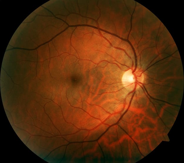
- The Silverstone RGB system includes nine imaging modalities for surface to deep tissue imaging, functional and metabolic imaging, vascular imaging, and structural cross-sectional imaging, significantly broadening diagnostic capabilities.
- It retains the ultra-widefield imaging and lesion-guided swept-source OCT from the existing Silverstone platform, ensuring continuity in clinical practice.
- Clinician feedback emphasized the need for greater access to peripheral pathology, intuitive modality switching, and faster image capturing, which were critical in shaping the system's development.
Interpretation:
The integrated approach of the Silverstone RGB system supports comprehensive pathology assessment, enhancing the ability of clinicians to make informed decisions based on a complete view of retinal health.
Limitations:
- The article does not provide specific data on the effectiveness or clinical outcomes of the Silverstone RGB system, which is crucial for evaluating its impact on patient care.
Conclusion:
The Silverstone RGB system aims to enhance retinal imaging capabilities while ensuring accessibility and efficiency in clinical practice, ultimately improving patient outcomes.
This content is an AI-generated, fully rewritten summary based on a published scholarly article. It does not reproduce the original text and is not a substitute for the original publication. Readers are encouraged to consult the source for full context, data, and methodology.