To identify specific brain network features associated with visual aura in migraine patients experiencing visual aura (VaM) compared to those without aura (MwoA) and healthy controls.
Approach:
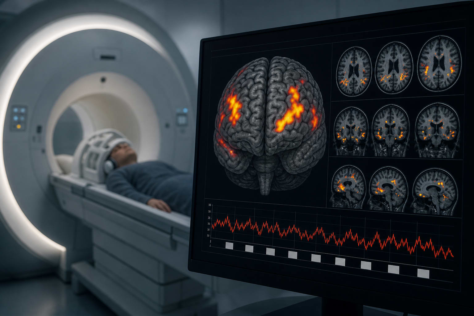
Participants: 42 migraine patients (21 VaM, 21 MwoA) and 21 healthy controls were recruited. All participants underwent visual stimulation with fMRI scanning.
Methods: Brain activation differences among the three groups were analyzed using one-way ANOVA and post-hoc comparisons. Within-group analyses were performed using one-sample t-tests.
Key Findings:
Differential activation was observed in multiple brain regions among the VaM, MwoA, and HC groups, including right cerebellar lobule IX, left middle temporal gyrus, and left orbital part of the inferior frontal gyrus.
VaM group showed stronger activation in the left precuneus compared to HC group.
MwoA group exhibited stronger activation in the right and left middle occipital gyrus compared to HC group.
VaM group demonstrated stronger activation in the right angular gyrus, left cuneus, and right middle cingulate gyrus compared to MwoA group.
VaM group showed weaker activation in the right middle occipital gyrus compared to MwoA group.
Interpretation:
Limitations:
The study was limited to a small sample size of 42 participants.
The findings are exploratory and require further investigation to confirm the mechanisms involved.
Conclusion:
The study identifies potential indicators of visual aura mechanisms in migraine patients.