Regional brain dysfunction patterns associated with rapid eye movement sleep behavior disorder and visual hallucinations in Parkinson’s disease: a resting-state fMRI study with exploratory ROI-based factorial analysis - Summary - MDSpire
Advertisement
Regional brain dysfunction patterns associated with rapid eye movement sleep behavior disorder and visual hallucinations in Parkinson’s disease: a resting-state fMRI study with exploratory ROI-based factorial analysis
To characterize regional brain dysfunction patterns associated with RBD and VH in PD and to explore candidate-region symptom-related effects within regions showing overall between-group differences.
Approach:
Study Design: Cross-sectional study involving 96 PD patients divided into four groups based on the presence or absence of RBD and VH.
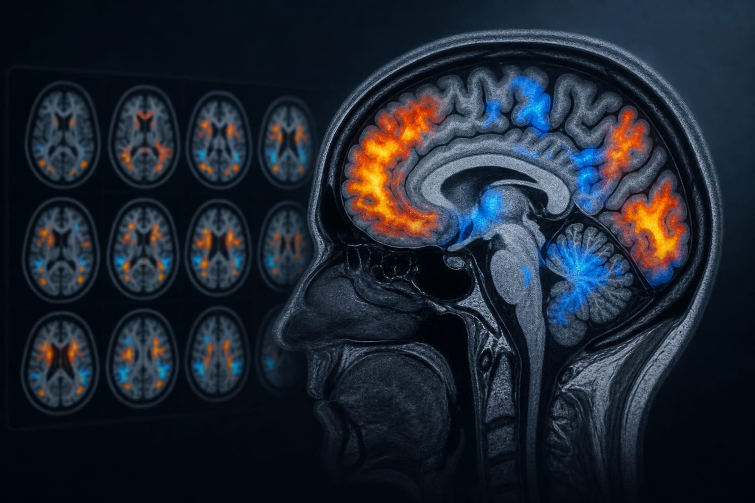
Imaging Analysis: Resting-state fMRI analyzed using amplitude of low-frequency fluctuations (ALFF) and regional homogeneity (ReHo).
Statistical Methods: Whole-brain four-group analyses followed by exploratory ROI-based 2 × 2 factorial analyses and voxel-wise analyses.
Key Findings:
Patients with both RBD and VH exhibited the greatest clinical burden and worse cognitive performance.
Whole-brain analyses revealed abnormalities in frontal, temporal, cerebellar, supplementary motor, and precuneus regions.
RBD-related patterns were found in precuneus ReHo, cerebellar lobule VIII ReHo, and SMA ALFF.
VH-related patterns were identified in OFC ReHo, precuneus ReHo, cerebellar Crus I ReHo, SMA ALFF, and temporal pole ALFF.
Imaging abnormalities correlated with RBD severity, freezing of gait, hallucination burden, and cognition.
Interpretation:
Coexisting RBD and VH may indicate a clinically more severe PD subtype associated with regional abnormalities in various brain regions.
Limitations:
Findings are exploratory and require confirmation in larger studies.
Imaging abnormalities were interpreted as post hoc exploratory results rather than independent confirmatory evidence.
Conclusion:
Symptom-related main and interaction patterns should be interpreted as candidate-region exploratory findings.
Aviva Abosch, M.D., Ph.D., a neurosurgeon at Baptist Health Miami Neuroscience Institute, part of Baptist Health Brain and Spine Care, was installed as the Esernia Endowed Chair in Surgical Treatment of Adult Epilepsy and Movement Disorders.