To investigate the long-term effects of LASIK on corneal nerves and immune cell behavior using advanced imaging techniques.
Approach:
Study Design: The study compared 13 post-LASIK patients with 19 age-matched healthy controls, evaluating corneal nerve architecture, sensory responses, tear cytokines, and immune cell behavior up to two years post-surgery.
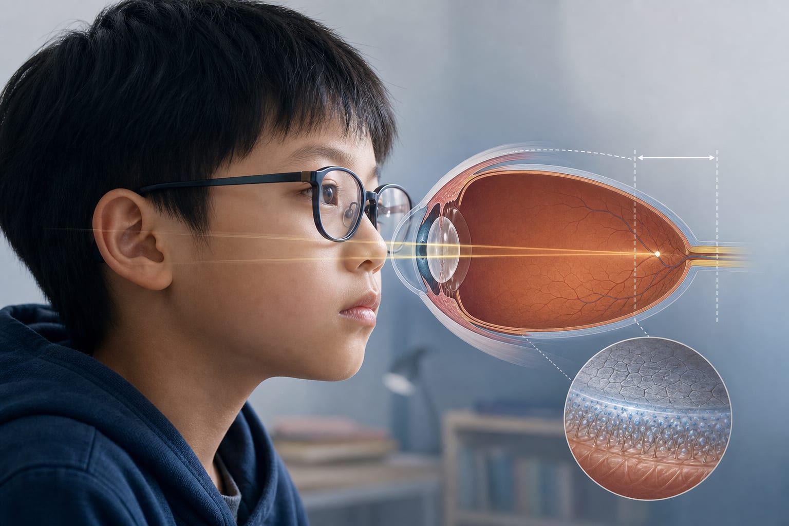
Imaging Technique: Advanced functional in vivo confocal microscopy (Fun-IVCM) was used to assess the ocular surface in detail.
Key Findings:
Post-LASIK corneas showed reduced central corneal nerve density and branching, along with impaired mechanical sensitivity.
Epithelial T cells in post-LASIK eyes exhibited significantly faster movement at the corneal whorl compared to controls, suggesting heightened immune surveillance activity.
Dendritic cells in post-LASIK participants had lower density but were larger, more circular, and morphologically distinct, indicating potential immune activation.
Elevated interleukin-16 (IL-16) levels were found in tears of post-LASIK patients, suggesting a prolonged low-grade inflammatory state.
Mechanical sensitivity was reduced, but responses to hyperosmolar saline stimuli were preserved.
Interpretation:
The study indicates that LASIK may induce persistent neuroimmune changes relevant for chronic post-LASIK dry eye and neuropathic ocular pain.
Limitations:
The clinical significance of the observed neuroimmune changes remains uncertain.
Subgroup analyses showed no major differences in immune cell morphology or dynamics between symptomatic and asymptomatic patients.
Conclusion:
Further studies are warranted to explore the long-term functional significance of the differences in corneal nerve and immune cell features after LASIK.