Primary central nervous system lymphoma and tumefactive demyelinating lesions in multiple sclerosis: a retrospective observational cohort study - Summary - MDSpire
Advertisement
Primary central nervous system lymphoma and tumefactive demyelinating lesions in multiple sclerosis: a retrospective observational cohort study
To compare the differences in clinical observations, neuroimaging data, and pathological features between primary central nervous system lymphoma (PCNSL) and tumefactive demyelinating lesions (TDLs), as well as their potential biomarkers.
Key Findings:
PCNSL patients had a mean onset age of 59 ± 10 years, while TDL patients had a mean age of 36 ± 8 years (p < 0.001).
MRI scans showed U-shaped hyperintense lesions in PCNSL and slightly diffuse hyperintense lesions in TDLs on DWI.
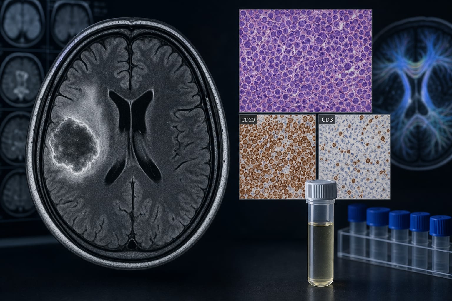
PCNSL lesions exhibited obvious hyperintense and perifocal edema on FLAIR sequences, whereas TDL lesions were isointense with partial perifocal edema.
CD20 was strongly positive in PCNSL tissues, while CD3 was negative; TDLs showed lower CD20 and stronger CD3 levels.
IL-6 and IL-10 levels were higher in the CSF of PCNSL patients compared to TDL patients.
Interpretation:
Remove or rephrase to avoid unsupported conclusions.
Limitations:
Add any limitations mentioned in the source.
Conclusion:
Revise to remove unsupported claims about the importance of evaluation.