Imaging markers for diabetic macular oedema: Forecasting visual outcomes beyond retinal thickness
-
By
-
Enrico Borrelli
-
October 18, 2026
Objective:
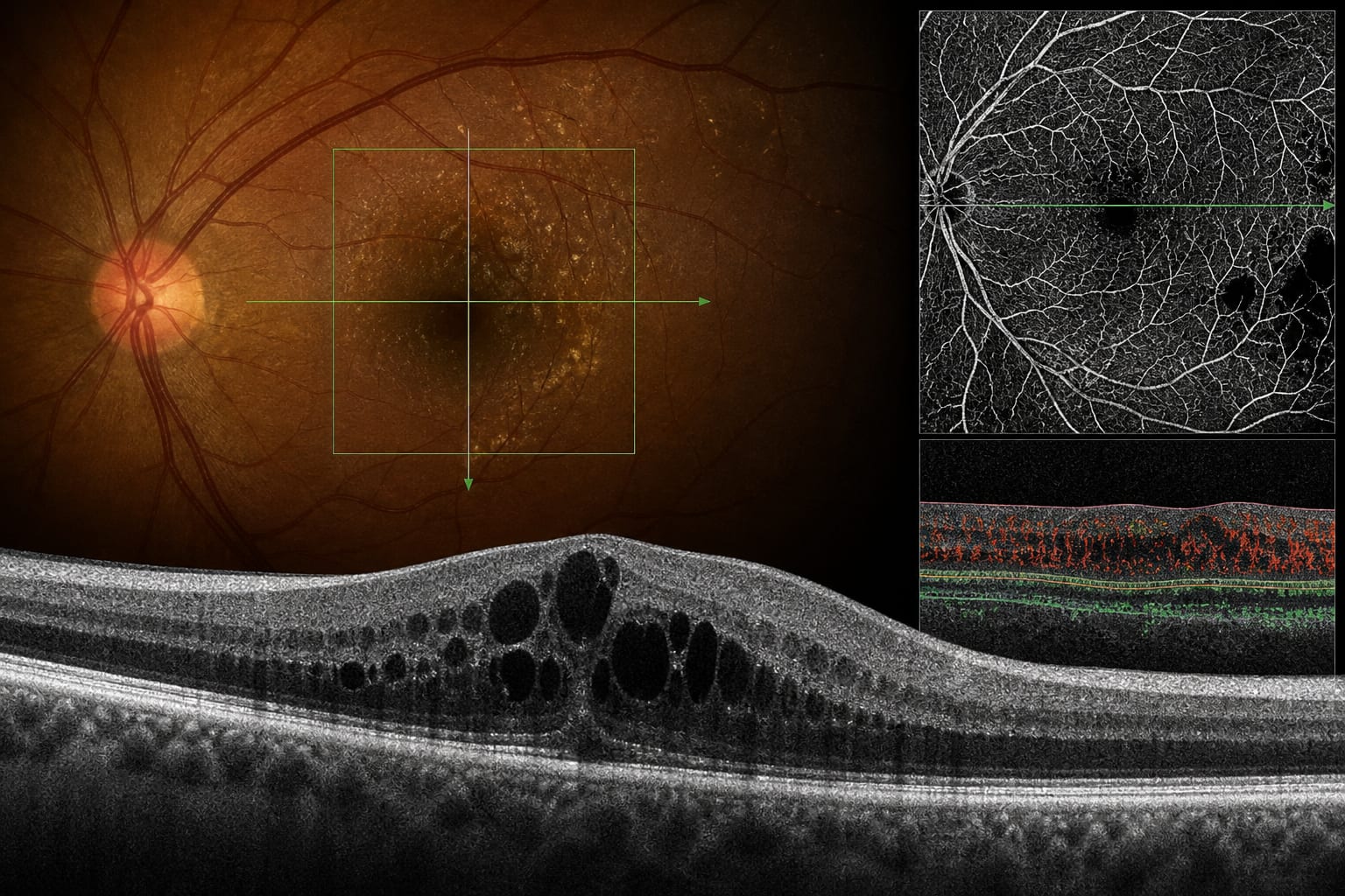
Summarise evidence on OCT and OCTA imaging markers associated with visual acuity and visual outcomes in diabetic macular oedema (DMO), including findings from Alhelaly et al.
Approach:
- Study: Post hoc analysis of 60 KINGFISHER trial eyes with available baseline spectral-domain OCT and OCTA measurements.
- Context: The article reviews structural OCT and OCTA biomarkers, prior evidence, and limitations affecting their validation and measurement.
Key Findings:
- Mean visual acuity in the analysed eyes increased from 62.9 letters at baseline to 75.4 letters at week 52.
- Greater baseline central subfield thickness (CSFT) independently predicted baseline visual acuity; baseline CSFT and a composite of superficial perfusion density and vessel length density independently associated with week-52 visual acuity.
- Higher baseline hyperreflective-foci volume was independently associated with a smaller visual-acuity gain.
- Prior evidence reviewed in the article links DRIL and outer-retinal alterations with visual outcomes; a meta-analysis of 27 studies found five biomarkers associated with reduced visual acuity at two or more time points, with moderate certainty.
- OCTA enables quantification of diabetic macular ischaemia across capillary plexuses using measures including perfusion density, vessel length density, and fractal dimension.
Interpretation:
In a post hoc analysis of 60 KINGFISHER trial eyes, baseline structural and vascular imaging measures were associated with visual acuity at baseline or week 52, while hyperreflective-foci volume was associated with visual-acuity gain. Alhelaly et al. describe diabetic macular ischaemia assessed by OCTA as one of the strongest predictors of long-term visual outcomes in anti-VEGF-treated DMO.
Limitations:
- The analysis included only 60 eyes, and OCTA was not required by the parent trial protocol.
- OCTA analysis was limited to scans obtained with the Heidelberg Spectralis.
- Models accounted for 19%–32% of variance and did not adjust for multiple comparisons.
- OCTA metrics vary with device, scan protocol, segmentation, artefact removal, and image-processing methods, limiting comparability across centres.
- The superficial vascular metrics were retained in models while deep-plexus metrics were excluded; this may reflect measurement consistency in oedematous eyes rather than greater biological relevance.
Conclusion:
The article calls for rigorous validation, standardised imaging acquisition and analysis, and prespecified imaging assessments in future clinical trials.
Sources:
Based on findings from:
Imaging biomarkers in diabetic macular oedema: Predicting vision beyond retinal thickness
Enrico Borrelli. Eye, 2026.
https://www.nature.com/articles/s41433-026-04986-0
This content is an AI-generated, fully rewritten summary based on a published scholarly article. It does not reproduce the original text and is not a substitute for the original publication. Readers are encouraged to consult the source for full context, data, and methodology.