Case report: Diffuse large B-cell lymphoma associated with chronic inflammation presenting as a rapidly enlarging perigraft hematoma after TEVAR - Summary - MDSpire
Advertisement
Case report: Diffuse large B-cell lymphoma associated with chronic inflammation presenting as a rapidly enlarging perigraft hematoma after TEVAR
To enhance clinicians’ awareness of diffuse large B-cell lymphoma associated with chronic inflammation (DLBCL-CI) in the context of postoperative perigraft masses, facilitating earlier diagnosis and reducing misdiagnosis.
Approach:
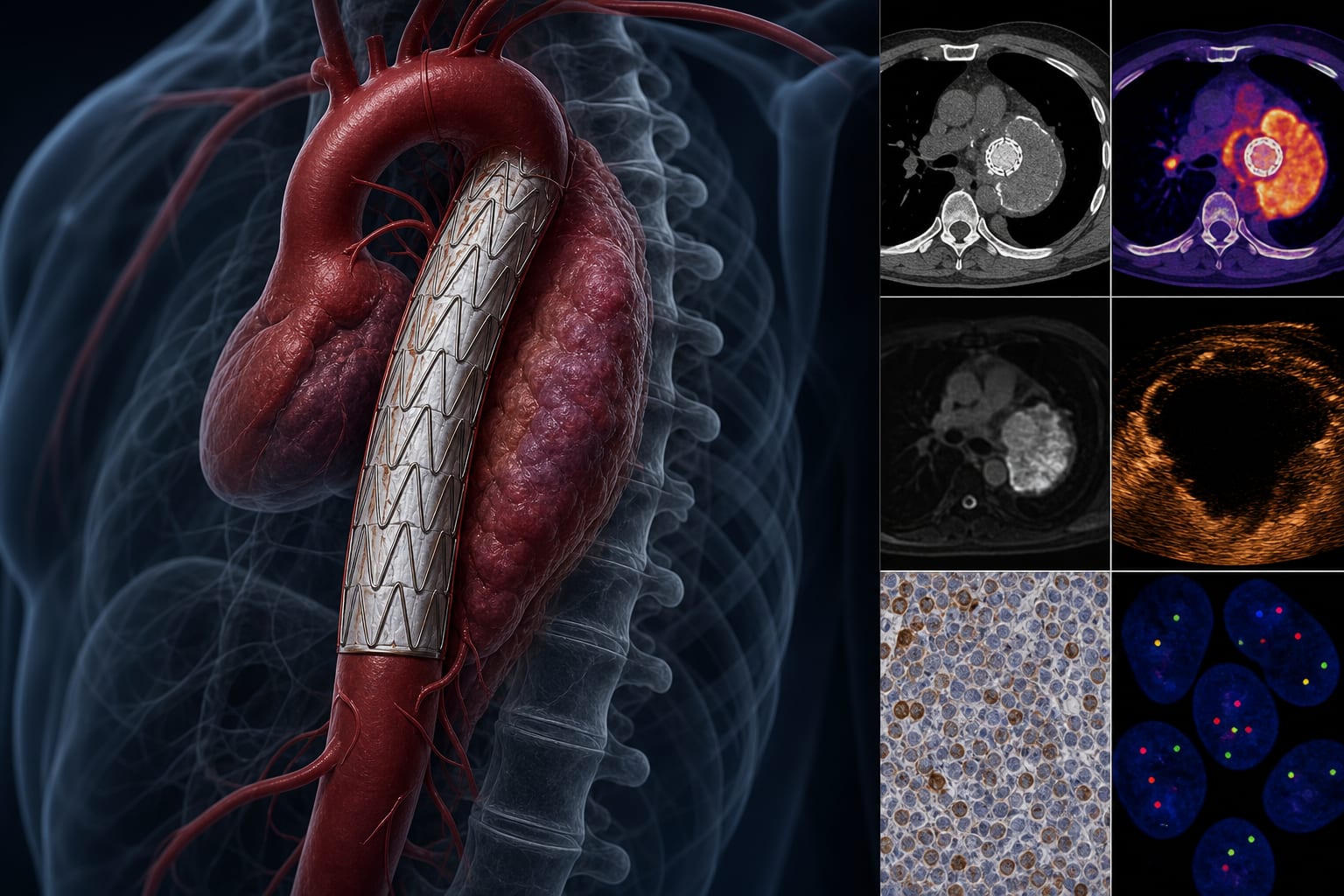
Case Presentation: A 59-year-old male underwent repeat TEVAR for stent-graft fracture. A periaortic hypodense lesion was initially interpreted as a hematoma but progressed into a large soft-tissue mass postoperatively.
Imaging Evaluation: Imaging included CTA, MRI, CEUS, and PET/CT, revealing discordant findings with restricted diffusion and absent enhancement, alongside increased metabolic activity.
Biopsy and Diagnosis: Core needle biopsy confirmed EBV-positive DLBCL with a TP53 mutation.
Key Findings:
The case represents a rare instance of DLBCL-CI around a vascular graft, meeting the criteria of a vascular graft background, EBER positivity, and formation of a well-defined mass.
This case is unique as it is one of fewer than five reported instances of DLBCL-CI occurring around vascular grafts.
Interpretation:
The findings highlight the need for heightened awareness of DLBCL-CI in the context of postoperative perigraft masses, as its early imaging features can overlap with those of hematoma or infection.
Limitations:
Limited number of reported cases of DLBCL-CI around vascular grafts, with fewer than five cases documented in the literature.
Potential for misdiagnosis due to overlapping imaging features with common postoperative complications.
Conclusion:
This case emphasizes the importance of considering DLBCL-CI in the differential diagnoses of perigraft masses following TEVAR, particularly given its rarity and the potential for misdiagnosis.