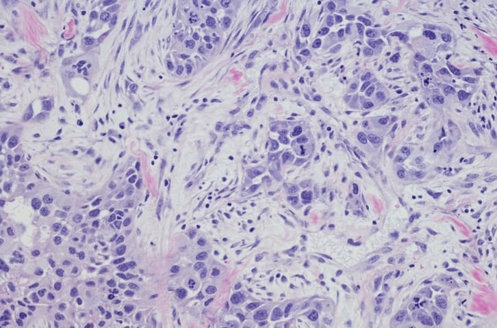
To describe variation in morphology, immunohistochemical findings, and clinical course in two patients with pulmonary NUT carcinoma.
Approach:
Cases: The authors reviewed tumor morphology, squamous differentiation, immunostaining, treatment, disease progression, and survival in two patients.
Diagnostic assessment: Testing included NUT, AE1/AE3, CAM5.2, p63, and p40 immunostaining, as well as Ki-67 labeling.
Key Findings:
One tumor showed prominent squamous differentiation and AE1/AE3 and CAM5.2 positivity; the other had only small foci of squamous differentiation, limited AE1/AE3 staining, and negative CAM5.2 staining.
Both tumors were positive for p63, p40, and NUT. Ki-67 labeling indices were approximately 30% and 50%.
Both patients developed aggressive disease progression and died: one 5 months after admission and the other 1 year after surgery.
The authors cited prior analyses reporting squamous differentiation in 33% of 124 NUT carcinoma cases and absent keratin expression in approximately 20%.
Interpretation:
The authors stated that inconspicuous squamous differentiation or negative keratin staining does not exclude pulmonary NUT carcinoma; diagnosis relies on positive NUT protein immunohistochemistry or molecular testing.
Limitations:
The report included only two patients and was descriptive, so it cannot establish how frequently these features occur.
Differences in tissue sampling and retrospective confirmation of the first diagnosis complicated direct comparison.
Conclusion:
The authors reported no funding or conflicts of interest.
In an exploratory subgroup analysis of patients with ESR1-mutated tumors, median progression-free survival was 11.1 months with the combination and 5.5 months with imlunestrant alone.