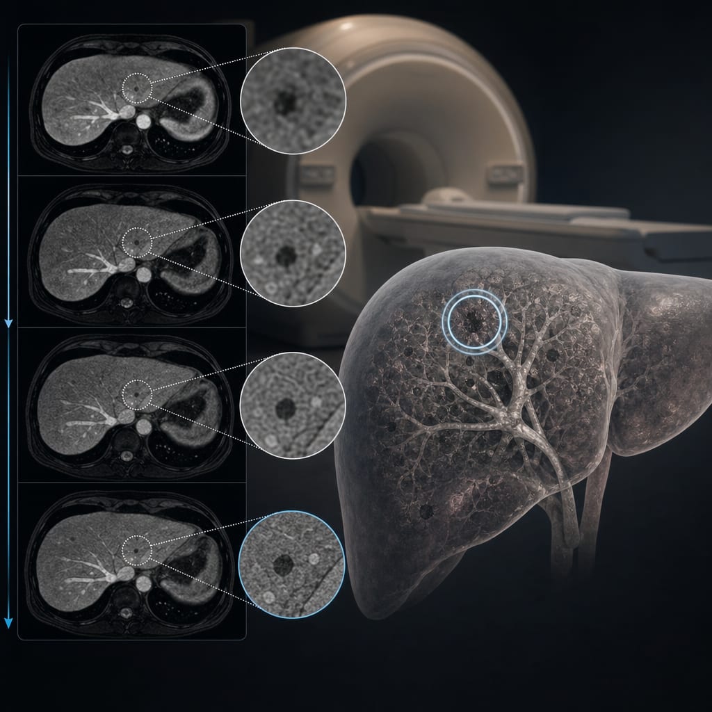
To evaluate the detection efficacy of Deep Learning Reconstruction (DL Recon) EOB-MRI against conventional Gd-EOB-DTPA-enhanced liver MRI during the hepatobiliary phase for identifying focal liver lesions (FLLs), highlighting the importance of accurate detection in clinical settings.
Key Findings:
Intraclass correlation coefficient (ICC) among all groups was >0.90, indicating excellent consistency (P < 0.001), suggesting high reliability in evaluations.
No significant differences in high-signal lesions among groups, except for Standard DL vs. Standard Non-DL (P = 0.048), indicating a potential advantage of DL in certain contexts.
For small low-signal lesions (5–10 mm), Standard DL showed significantly more lesions than Standard Non-DL (P = 0.030), emphasizing its effectiveness in detecting smaller lesions.
Both DL and Non-DL HR methods revealed significantly greater numbers of small low-signal lesions (<5 mm) compared to Non-DL alone (P ≤ 0.032), underscoring the enhanced detection capabilities.
Interpretation:
DL-Recon significantly enhances the detection of small low-signal abnormalities in EOB-MRI hepatobiliary phase, improving the identification rate of FLLs.
Limitations:
Single-center study may limit generalizability; further multi-center studies are needed to validate findings.
Sample size of 53 patients may not be sufficient for broader conclusions; larger studies are recommended to confirm results.
Conclusion:
DL-Recon integrated with MR methodologies shows considerable clinical importance in boosting the identification of diminutive low-signal abnormalities in EOB-MRI hepatobiliary phase, significantly enhancing the detection rate of FLLs and potentially impacting patient management.