To investigate the cellular and molecular mechanisms driving radiation-induced heart injury (RIHI) using single-cell RNA sequencing.
Approach:
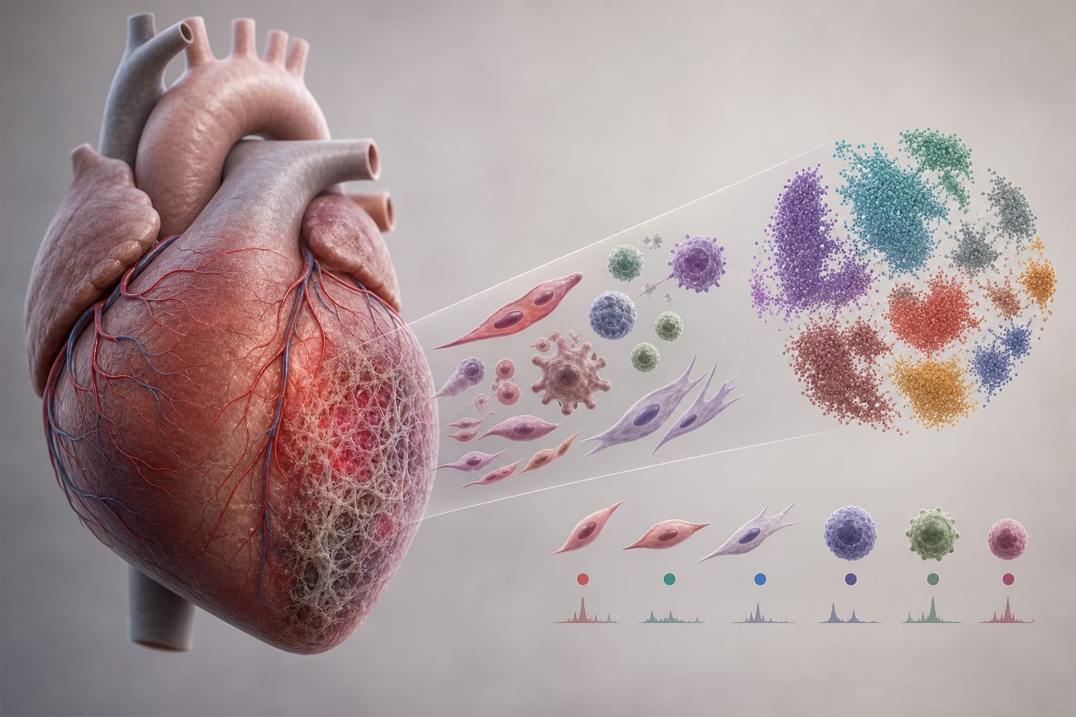
Single-cell RNA sequencing: Performed on rat hearts and matched peripheral blood mononuclear cells (PBMCs) 12 weeks post whole-heart irradiation (20 Gy) or sham control, profiling 38,941 cardiac cells and 41,097 PBMCs.
Analytical methods: Conducted differential expression, pathway enrichment, pseudotime, and ligand-receptor interaction analyses; validated key findings using Western blotting and flow cytometry.
Key Findings:
Defined major cardiac populations, including cardiomyocytes, endothelial cells, fibroblasts, and various immune cells in control and RIHI hearts.
Endothelial cells exhibited subtype shifts and marked MHC-II upregulation post-irradiation.
Fibroblasts showed iron accumulation and pro-inflammatory activation with antigen-presenting properties.
Myeloid activation and T/NK cell polarization toward cytotoxic but partially exhausted states were observed.
Enhanced B-cell antigen presentation was noted, linking radiation injury to chronic cardiac inflammation.
Interpretation:
RIHI progresses through a stromal-immune cascade where endothelial and fibroblast immunogenic reprogramming initiates sustained myeloid and lymphoid activation, creating a pro-inflammatory cardiac microenvironment.
Limitations:
Study conducted in a rat model, which may not fully replicate human responses to radiation.
Focus on specific time points post-irradiation may overlook earlier cellular changes.
Conclusion:
Findings suggest non-hematopoietic antigen presentation as a potential therapeutic target in thoracic radiotherapy, especially in combination with immune checkpoint inhibitors.