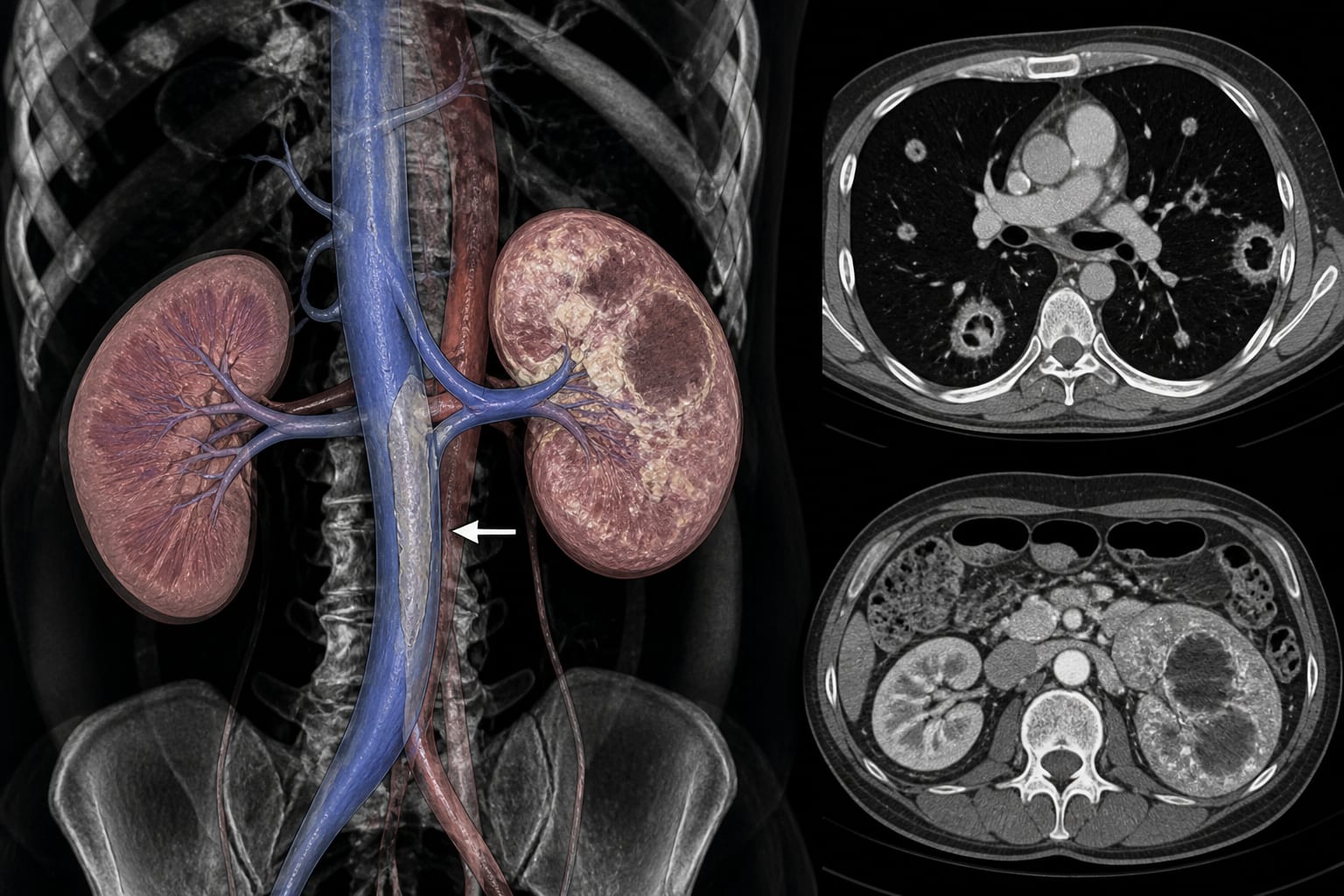
To report a case of presumed hypervirulent Klebsiella pneumoniae pyelonephritis causing renal vein–inferior vena cava septic thrombophlebitis, cavitary septic pulmonary emboli, and lung abscesses in a patient with untreated diabetes.
Approach:
Patient Presentation: A 47-year-old woman with untreated diabetes (hemoglobin A1c, 14.7%) presented with dysuria, 11 days of fever, abdominal pain, and hypoxemia.
Microbiological Findings: Blood and urine cultures grew K pneumoniae. Bacteremia persisted despite meropenem. The isolate was susceptible to ceftriaxone and levofloxacin and produced an 11-mm positive string test, supporting a hypermucoviscous phenotype.
Imaging and Cardiac Evaluation: Contrast-enhanced CT demonstrated left pyelonephritis, renal abscesses, a near-occlusive thrombus extending from the left renal vein into the inferior vena cava, bilateral peripheral cavitary pulmonary nodules, and pulmonary arterial emboli. Transesophageal echocardiography showed no vegetation.
Treatment and Management: Therapy was changed to ceftriaxone at meningitis dosing plus levofloxacin, with anticoagulation and insulin treatment. Nephrectomy, thrombectomy, and inferior vena cava filter placement were considered hazardous.
Clinical Course: Blood cultures cleared by day 7. Pulmonary lesions worsened on day 10, but clinical, laboratory, and radiographic improvement by day 38 supported continued medical management without intervention.
Key Findings:
K pneumoniae pyelonephritis with renal abscesses was associated with renal vein–inferior vena cava septic thrombophlebitis and cavitary septic pulmonary emboli.
The positive string test supported a hypermucoviscous phenotype, but hypervirulence remained presumptive because genotyping was unavailable.
Persistent bacteremia despite meropenem and negative transesophageal echocardiography prompted evaluation for an occult septic venous source.
Continued antimicrobial therapy, anticoagulation, and insulin treatment produced regression of the infected thrombus and pulmonary lesions.
Antimicrobials were discontinued on day 49. By day 117, the thrombus and septic pulmonary emboli had nearly resolved, allowing rivaroxaban to be discontinued.
Interpretation:
Persistent K pneumoniae bacteremia accompanied by peripheral cavitary pulmonary nodules should prompt consideration of venous imaging for occult septic thrombophlebitis, particularly in patients with urinary infection, renal abscesses, diabetes, or negative echocardiography. This case also illustrates that early radiographic progression may occur despite subsequent clinical improvement, although management decisions must remain individualized.
Limitations:
Genotyping was unavailable, so the isolate’s hypervirulence could not be genetically confirmed despite its hypermucoviscous phenotype.
This report describes a single patient and cannot establish the frequency, causality, or comparative effectiveness of the management approach.
Conclusion:
Medical treatment with ceftriaxone, levofloxacin, anticoagulation, and insulin was followed by clearance of bacteremia and near-complete resolution of the renal vein–inferior vena cava thrombus and septic pulmonary emboli without surgical intervention. More than 4 years after antimicrobial therapy ended, no infection, thrombosis, or septic pulmonary embolism had recurred.