Atrial volume reduction correlates with early improvement in hemorrhage-associated normal pressure hydrocephalus—a 3D computed tomography volumetric study - Summary - MDSpire
Advertisement
Atrial volume reduction correlates with early improvement in hemorrhage-associated normal pressure hydrocephalus—a 3D computed tomography volumetric study
To determine the relationship between changes in ventricular volume and early clinical improvement in patients with hemorrhage-associated normal pressure hydrocephalus (HANPH) after ventriculoperitoneal shunting (VPS).
Approach:
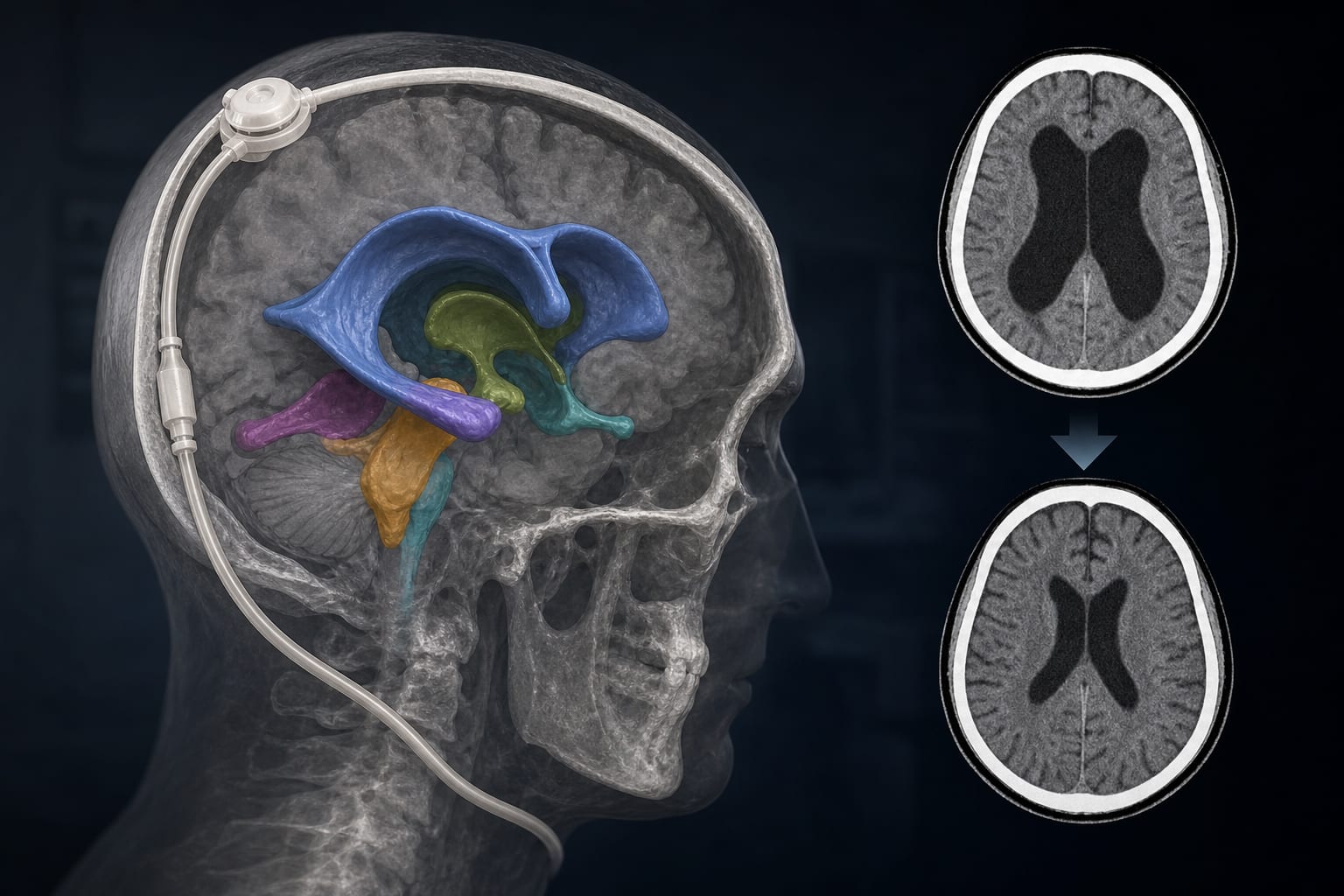
Study Design: Retrospective pre-post within-subject study involving 180 adult HANPH patients who underwent VPS.
Data Collection: CT scans were obtained preoperatively and 2 weeks post-VPS, measuring lateral ventricular subregions using 3D Slicer.
Clinical Assessment: Gait ability, modified Rankin Scale (mRS), and Barthel Index (BI) were assessed to evaluate clinical improvement.
Key Findings:
Significant reductions in absolute volumes of lateral ventricular subregions were observed post-VPS.
Postoperative relative volume of the lateral body of the lateral ventricle increased, while other subregions decreased.
Reductions in the absolute volumes of the frontal horn, temporal horn, and atrium negatively correlated with early clinical improvement.
Logistic regression revealed that for every 5‰ reduction in volume of these subregions, the likelihood of clinical improvement significantly increased by 180.3%, 340.9%, and 504.2%, respectively.
Interpretation:
The reduction in atrial volume of the lateral ventricle is crucial for early clinical improvement in HANPH post-VPS.
Limitations:
Retrospective nature of the study may introduce bias.
Lack of randomization in group assignment.
Conclusion:
Catheter placement in the occipital horn may optimize outcomes by effectively reducing atrial volume and minimizing risks.