Machine learning combined with resting-state functional MRI to characterize functional brain differences in post-stroke depression - Summary - MDSpire

Machine learning combined with resting-state functional MRI to characterize functional brain differences in post-stroke depression

  • By

  • Yuanxin Shao

  • Chao Liang

  • Dan Xu

  • Yang Zhao

  • Phi Thi Thanh Hoa

  • Xue Zhang

  • Dongyang Shi

  • Weifeng Guo

  • June 22, 2026

  • 0 min

Share

Objective:

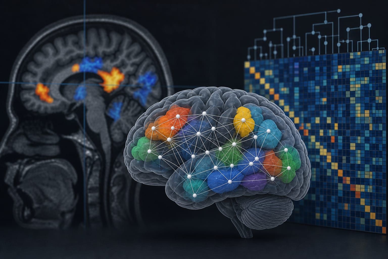
To examine resting-state functional differences between patients with post-stroke depression (PSD) and healthy controls, and to evaluate the use of machine learning to identify imaging features associated with PSD.
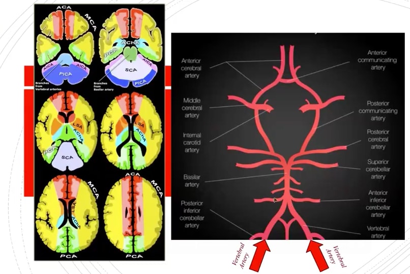
Approach:
    Key Findings:
    • PatientswithPSDshowedresting-statefunctionaldifferencesincingulate,thalamic,prefrontal,insular,posteriordefault-mode,andvisual-associatedregions.Twenty-ninecandidatefeaturesdifferedbetweengroups,including7ReHo,8ALFF,6DC,and8FCfeatures.LASSOretained10corefeatures,withacross-validatedAUCof0.878.TheExtraTreesmodelachievedthehighestindependenttest-setperformancewithanAUCof0.889.KeyinfluentialfeaturesincludedDCintheleftanteriorcingulate,ReHointheleftthalamus,andFCbetweentheleftprecuneusandleftcalcarinecortex.
    Interpretation:

    The study identified multi-level resting-state functional differences in PSD, with machine learning revealing candidate rs-fMRI features.

    Limitations:
    • Findings require validation in larger longitudinal cohorts.
    • The specificity and clinical utility of identified features need further clarification.
    Conclusion:

    The study highlights the integration of machine learning with rs-fMRI to understand PSD-related brain function variations.

Original Source(s)

Related Content