To present a case of anomalous origin of the left anterior descending artery from the pulmonary artery (ALADAPA) and highlight the importance of multimodality imaging in diagnosis.
Approach:
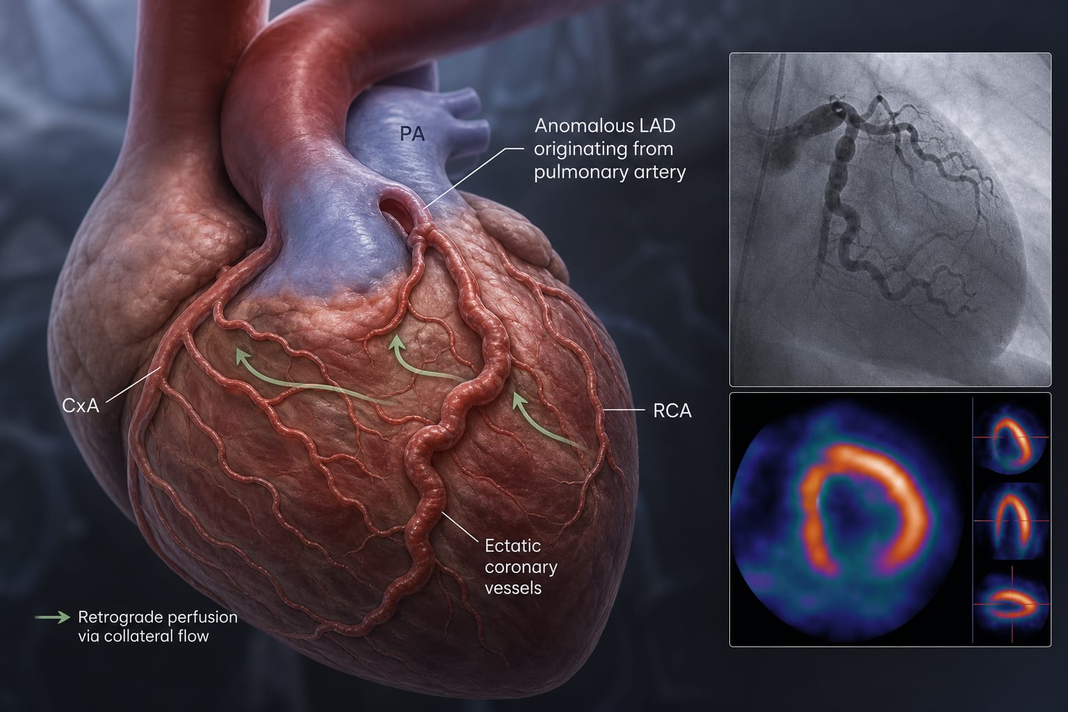
Case Presentation: A 59-year-old woman with progressive dyspnea underwent echocardiography, coronary angiography, and CT angiography, revealing ectatic coronary arteries and ALADAPA.
Diagnostic Imaging: Transthoracic echocardiography showed left ventricle dilation and diastolic flow signals. Coronary angiography confirmed the anomalous artery origin, and CT angiography supported these findings.
Functional Assessment: Myocardial perfusion scintigraphy indicated moderate-to-severe reversible ischemia in the anterior myocardial territory.
Surgical Intervention: The patient was referred for surgical correction to restore antegrade perfusion.
Key Findings:
ALADAPA is exceptionally rare, with only 52 reported cases.
The condition may remain undetected until adulthood due to collateral development.
Multimodality imaging is crucial for early recognition and diagnosis.
Interpretation:
Surgical reimplantation of the anomalous artery into the aorta is a treatment option to address complications such as myocardial ischemia and sudden cardiac death.
Limitations:
The rarity of ALADAPA may limit the generalizability of findings to broader populations.
Only a few cases have been documented in the literature, which may affect the understanding of the condition.
Conclusion:
Recognizing ALADAPA early and considering surgical intervention may help manage the condition and reduce the risk of serious cardiac events.
A review of cardiogenic shock studies suggests serial lactate measurements provide more prognostic information than isolated values and may better reflect treatment response.