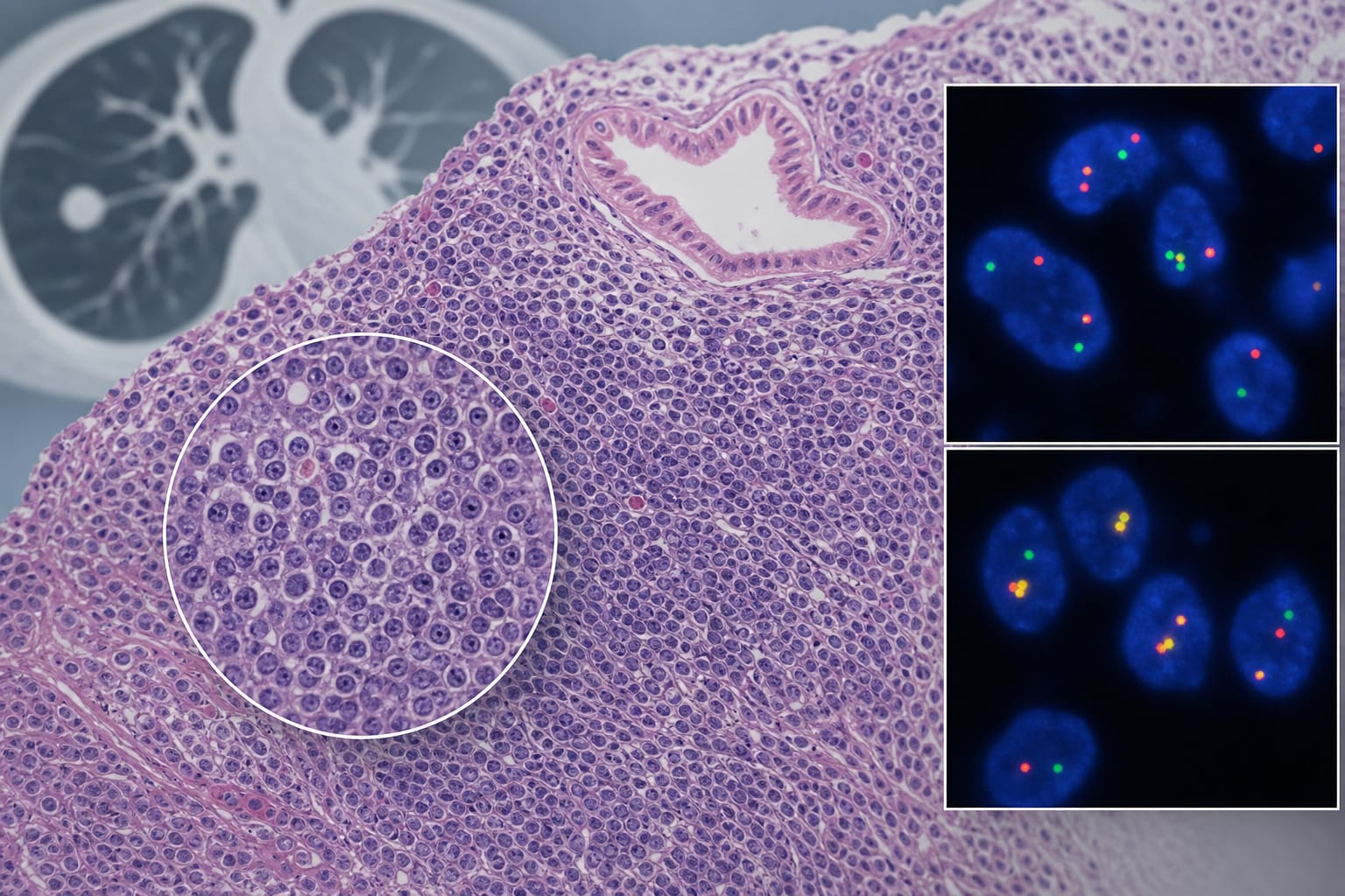
To present a case of pulmonary MALT lymphoma with BIRC3::MALT1 fusion.
Approach:
Case Presentation: A 49-year-old man underwent surgery for a pulmonary nodule, diagnosed with pulmonary MALT lymphoma, and found to have a BIRC3::MALT1 fusion.
Diagnostic Assessment: Histopathology, immunohistochemistry, and fluorescence in situ hybridization were used to confirm the diagnosis and assess disease extent.
Imaging Review: Longitudinal imaging and retrospective review of CT scans were conducted to evaluate the stability of pulmonary opacities.
Key Findings:
The patient had a confirmed diagnosis of pulmonary MALT lymphoma with a BIRC3::MALT1 fusion.
Postoperative PET/CT showed mild FDG uptake in longstanding pulmonary opacities and an ambiguous gastrointestinal finding.
Longitudinal imaging indicated that the right-lung ground-glass opacities were stable and predated the surgery.
Interpretation:
Mild FDG uptake in longstanding pulmonary opacities and unbiopsied gastrointestinal abnormalities should not be interpreted as lymphoma dissemination.
Limitations:
The study is based on a single case, limiting generalizability.
Ambiguous PET/CT results may lead to misinterpretation of disease extent.
Conclusion:
Integration of histopathology, immunophenotyping, molecular testing, and longitudinal imaging is essential before assigning disease extent.