To compare the diagnostic accuracy of FAP-targeted imaging (68Ga-FAPI) with the established imaging standard (18F-DOPA) for the detection of metastases in patients with medullary thyroid carcinoma (MTC).
Approach:
Study Design: A retrospective study comparing 18F-DOPA and 68Ga-FAPI PET/CT imaging using per-patient and per-lesion analyses in patients with recurrent MTC.
Patient Inclusion: Nine patients with histologically confirmed MTC who underwent both imaging modalities within a 3-month period were included.
Imaging Analysis: Imaging findings were correlated with morphological imaging or histopathology as the gold standard, and quantitative assessments included standardized uptake values (SUVs) and tumor-to-background ratios (TBRs).
Key Findings:
68Ga-FAPI showed significantly higher lesion detection rates for lymph node metastases (TP rate: 100% [8/8] vs. 50% [4/8] for 18F-DOPA).
For lung metastases, 68Ga-FAPI had a TP rate of 85.7% [12/14] compared to 42.9% [6/14] for 18F-DOPA.
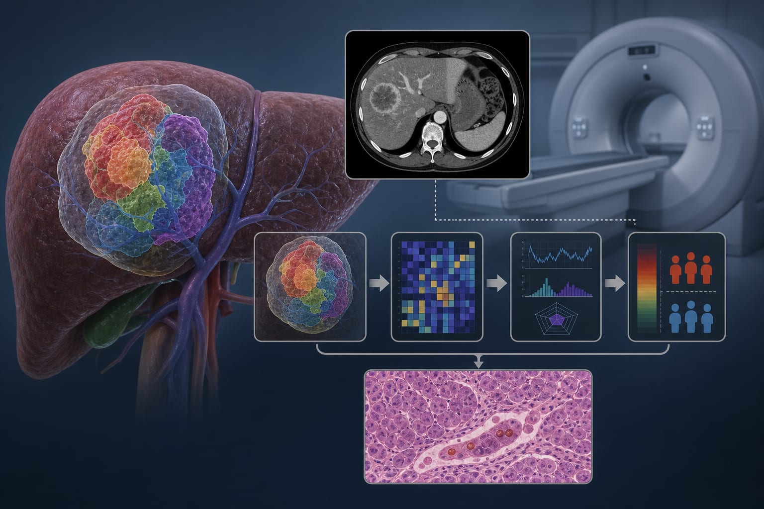
In liver metastases, 68Ga-FAPI detected 100% [22/22] of lesions versus 23% [5/22] for 18F-DOPA.
Higher TBR values were observed with 68Ga-FAPI compared to 18F-DOPA.
Interpretation:
68Ga-FAPI PET/CT detected a higher number of metastases with improved TBR values compared to 18F-DOPA.
Limitations:
The study included a small sample size of only nine patients.
Findings require confirmation in a larger, prospective study.
Conclusion:
Further validation of 68Ga-FAPI PET/CT is necessary.
by Daniel A. Hescheler, Dogan Atiyat, Kerstin Lorenz, Alexander Heinzel, Philipp Reschke, Frankis G. Almaguel, Manuela Petersen, Elisabeth Eppard, Joanna Wybranska, Jan Wuestemann, Michael C. Kreissl