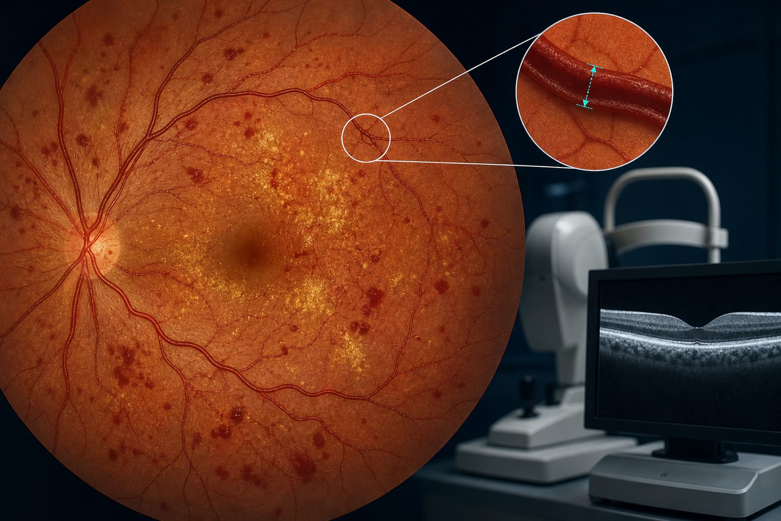
To detect GDF15 levels in aqueous humor (AH) and vitreous humor (VH) of patients with proliferative diabetic retinopathy (PDR) and analyze its differential expression.
Approach:
Sample Collection: AH and VH samples were collected from PDR patients and compared with controls from cataract patients and those with macular hole or epiretinal membrane.
Quantification Method: GDF15 levels were quantified using enzyme-linked immunosorbent assay (ELISA).
Key Findings:
GDF15 levels in AH of PDR patients were significantly higher than those in cataract controls.
GDF15 levels in VH of PDR patients were markedly higher than those in macular hole/epiretinal membrane controls.
An exploratory comparison showed higher GDF15 levels in VH than in AH in independent PDR patient cohorts.
Interpretation:
GDF15 expression is significantly upregulated in AH and VH of PDR patients.
Limitations:
The study involved a small sample size.
Further research is needed to elucidate the mechanisms of GDF15 in the intraocular microenvironment.
Conclusion:
The findings provide evidence for the role of GDF15 in the intraocular microenvironment of PDR.