To highlight the challenges in detecting wooden foreign bodies in clinical imaging studies.
Approach:
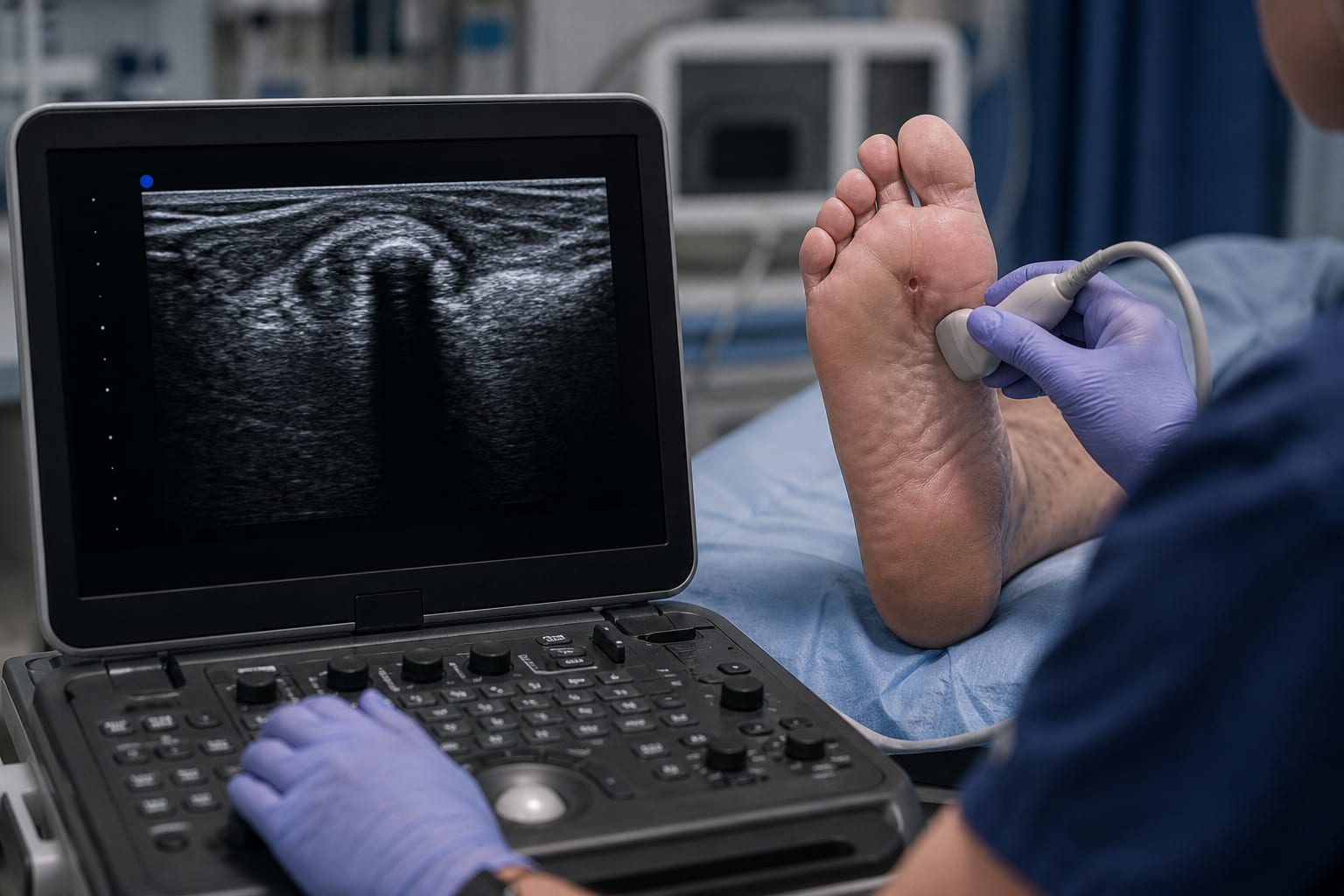
Case Description: A 69-year-old man with a history of diabetes presented with severe left foot pain after stepping on a sharp object. Upon examination, marked erythema and 2+ pitting edema were observed in the left foot, extending proximally to the tibial tuberosity. A 1.5 mm puncture wound without drainage was noted on the plantar aspect, specifically near the second metatarsophalangeal joint. Despite comprehensive imaging studies, including CT and MRI, which failed to identify any foreign body, point-of-care ultrasound was performed and revealed a prominent vertical shadow at the puncture site, raising suspicion for a retained foreign body. Subsequent surgical wound exploration confirmed the presence of a 1.8 cm wooden toothpick.
Imaging Modalities: Plain radiographs are capable of detecting approximately 80% of foreign bodies; however, wooden objects are often undetectable in a significant number of cases due to their composition. CT scans are not optimal for detecting wooden foreign bodies because the density of wood closely resembles that of soft tissues, leading to potential misinterpretation. MRI also has limited sensitivity in identifying wooden materials. In contrast, ultrasound has proven to be an effective tool for identifying wooden foreign bodies, especially those that are lodged within superficial soft tissues, owing to its ability to visualize acoustic shadows created by such materials.
Key Findings:
Ultrasound is the preferred imaging modality for detecting wooden foreign bodies due to its high sensitivity and ability to visualize soft tissue structures.
CT and MRI are less effective for identifying wooden materials, which can lead to missed diagnoses.
Interpretation:
Given the findings, point-of-care ultrasound should be considered a primary imaging technique for suspected retained non-metallic foreign bodies, as it demonstrates superior sensitivity compared to CT and MRI.
Limitations:
The study is based on a single case report, which may limit the generalizability of the findings to broader clinical populations.
Data availability is restricted due to privacy or ethical concerns, which may affect the comprehensiveness of the analysis.
Conclusion:
Ultrasound is an effective imaging modality for detecting wooden foreign bodies in clinical settings, highlighting the need for its consideration in cases of suspected non-metallic foreign body retention.