Evaluation of intraoperative color Doppler ultrasonography in the surgical evacuation of acute intracranial hematoma: a single-center study from Botswana - Summary - MDSpire
Advertisement
Evaluation of intraoperative color Doppler ultrasonography in the surgical evacuation of acute intracranial hematoma: a single-center study from Botswana
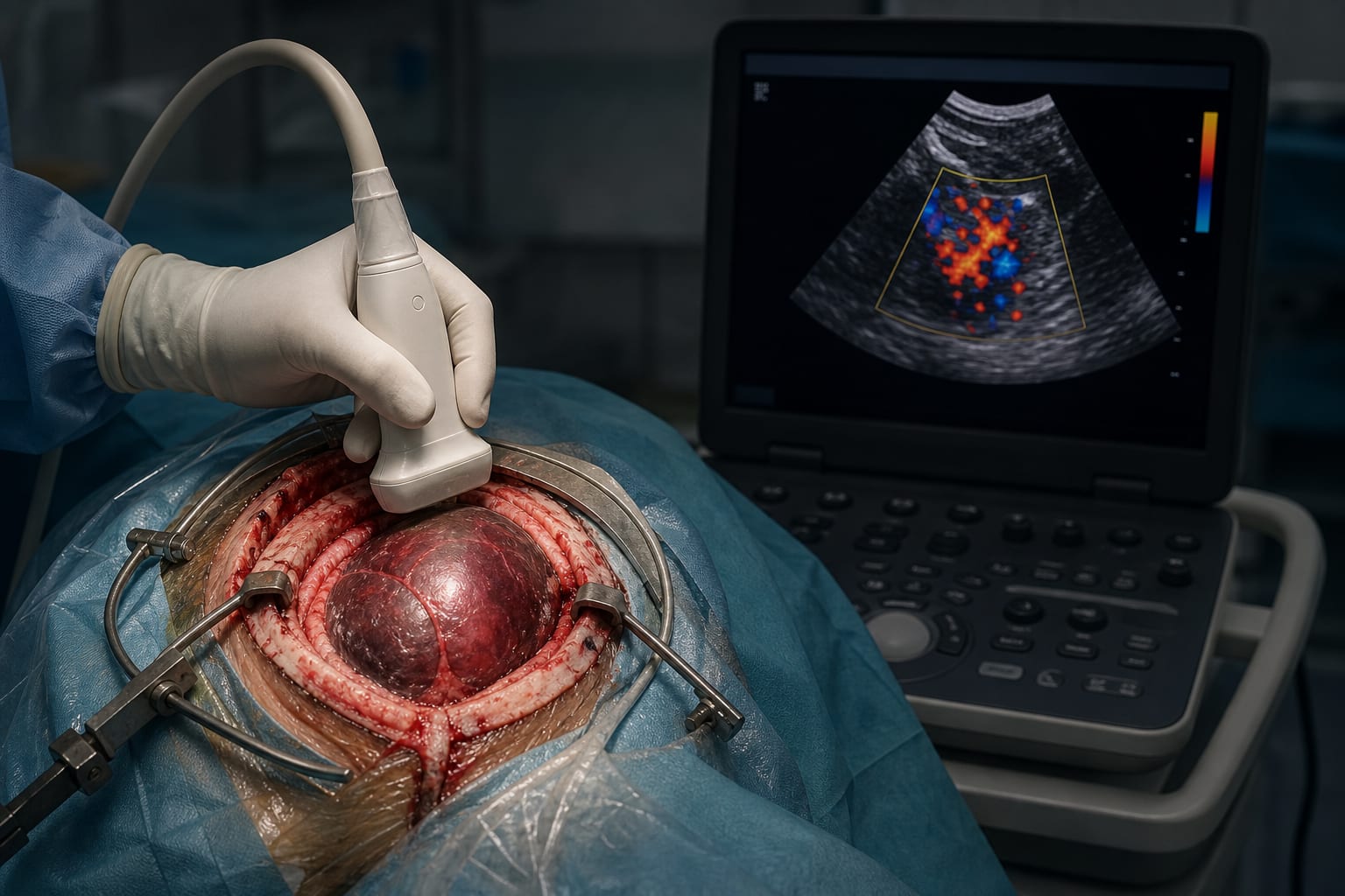
To evaluate the effectiveness of intraoperative color Doppler ultrasonography in improving hematoma clearance and neurological outcomes in patients undergoing craniotomy for acute intracranial hemorrhage, particularly in resource-limited environments, highlighting its significance.
Key Findings:
70.6% of the ultrasound-guided group achieved >90% hematoma clearance compared to 26.1% in the control group (P < 0.001).
The ultrasound-guided group had higher GCS scores (12 vs. 9, P < 0.001) and lower NIHSS scores (8.38 vs. 11.78, P < 0.001) two weeks postoperatively.
Shorter hospital stay for the ultrasound-guided group (12.47 days vs. 16.39 days, P = 0.024).
Intraoperative ultrasonography detected residual, newly formed, and contralateral hematomas not visible on preoperative CT.
Interpretation:
Intraoperative color Doppler ultrasonography provides real-time, dynamic information that enhances surgical clearance rates and improves short-term neurological outcomes in patients with acute supratentorial hematomas, with significant implications for clinical practice.
Limitations:
Single-center study may limit generalizability and affect the robustness of findings.
Small sample size may affect the robustness of findings.
Exclusion of certain hemorrhage types limits applicability and may skew results.
Conclusion:
Intraoperative color Doppler ultrasonography is a practical adjunct to CT-guided neurosurgery, particularly beneficial in resource-constrained settings, enhancing surgical outcomes for acute intracranial hematomas and potentially improving patient outcomes.