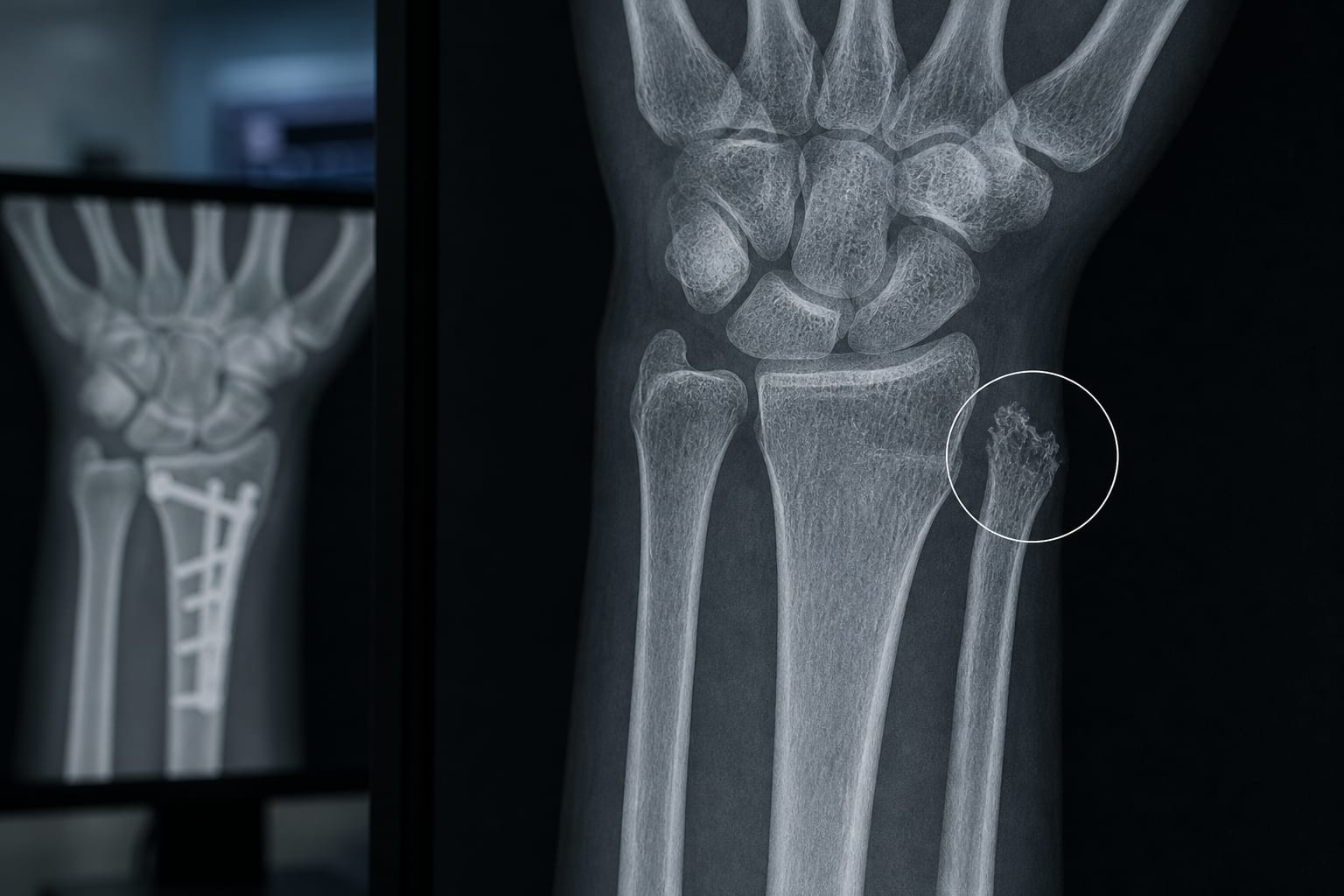
The article examines the objective, findings, and clinical context described in Ulnar Styloid Osteolysis: A Clinically Meaningful Finding Beyond Imaging?.
Approach:
Study design: Retrospective analysis of 143 individuals with distal radius fractures.
Assessment: Follow-up radiographs were used to identify ulnar styloid osteolysis and examine associated factors.
Key Findings:
Ulnar styloid osteolysis was identified in 16.8% of cases and usually became apparent during the first months after injury.
Osteolysis was associated with fracture comminution, dorsal angulation, diastasis, impaction, and active smoking.
The study characterizes osteolysis as a post-traumatic imaging finding, but its biological basis and relationship to wrist function remain uncertain.
Interpretation:
The finding may be relevant to radiographic differential diagnosis after distal radius fracture. Whether it reflects true cortical bone loss, insertional remodeling, occult microfracture, or another process—and whether it relates to TFCC injury or DRUJ dysfunction—has not been established.
Limitations:
The retrospective study relied almost entirely on follow-up radiographs, limiting characterization of the lesions’ structural basis.
The available evidence does not establish whether osteolysis has measurable clinical consequences.
Conclusion:
The study reports ulnar styloid osteolysis as a relatively frequent post-traumatic observation and provides a basis for further investigation. The article identifies CT and MRI as potential tools for clarifying lesion structure and relationships with DRUJ instability and TFCC pathology.