To highlight the role of advanced MRI techniques in the diagnosis, treatment planning, and monitoring of high-grade gliomas, ultimately aiming to improve patient outcomes.
Key Findings:
High-grade gliomas are aggressive tumors with poor prognosis despite treatment advancements, necessitating improved imaging techniques.
MRI is essential for glioma management, providing superior soft tissue contrast and advanced imaging capabilities that inform treatment decisions.
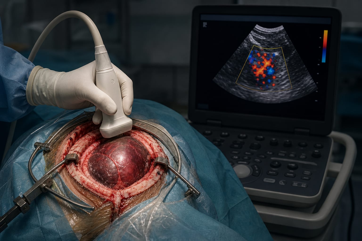
Perfusion MRI techniques, particularly DSC, DCE, and ASL, offer valuable insights into tumor vascularity and behavior, impacting clinical outcomes.
DWI and DTI are critical for understanding tumor microstructure and aiding in surgical interventions, influencing patient management strategies.
Interpretation:
The integration of advanced MRI techniques is crucial for improving the management of high-grade gliomas, allowing for better characterization, treatment planning, and ultimately enhancing patient outcomes.
Limitations:
DSC is sensitive to artifacts and may be inaccurate in certain conditions, which can affect treatment planning.
DCE has lower signal-to-noise ratio and requires complex post-processing, potentially complicating clinical use.
ASL is sensitive to motion and has lower spatial resolution, which may limit its effectiveness in certain patient populations.
Conclusion:
Advanced MRI techniques enhance the understanding and management of high-grade gliomas, but the choice of method should consider clinical context, patient-specific factors, and the potential impact on treatment outcomes.